Figure 3. Immnostaining of pannus tissue against vimentin and collagen IV. Compared to control (C, normal cornea), collagen IV expression (red) was irregular in pannus, and was also found in vessel walls (A and B). Vimentin(+) (green) stromal cells were uniformly observed in normal central cornea, but supra- and sub-epithelial activated
vimentin(+) cells were observed in pannus. Cells double-positive for vimentin and p63 were observed (D, indicated by arrow). The scale bar indicates 50 μm.
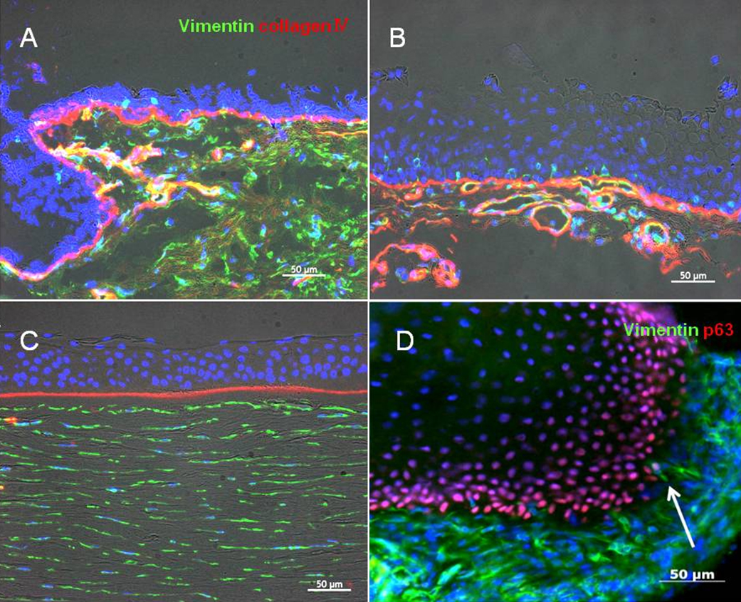
 Figure 3 of
Kawashima, Mol Vis 2010; 16:2727-2732.
Figure 3 of
Kawashima, Mol Vis 2010; 16:2727-2732.