Figure 2. Immunostaining of pannus tissue against pancytokeratin (CK22). Epithelium of pannus in all cases stained positive for pancytokeratin
(red), particularly in superior hyperepithelial layers. However, basal cells showed weak staining (A and B). C: pancytokeratin staining in normal cornea as a control. The scale bar indicates 50 μm.
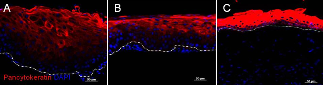
 Figure 2 of
Kawashima, Mol Vis 2010; 16:2727-2732.
Figure 2 of
Kawashima, Mol Vis 2010; 16:2727-2732.