Figure 1. Clinical photos of polypoidal choroidal vasculopathy (PCV). A: Color fundus photograph of a patient with PCV. Several orange lesions are visible in the macula. Hemorrhage is visible between
the orange lesions and the optic disk. B: The diagnosis of PCV was confirmed with indocyanine green angiography (ICGA). Abnormal choroidal vascular networks and characteristic
polypoidal lesions are visible in the macula, corresponding to the orange lesion in the color fundus photograph.
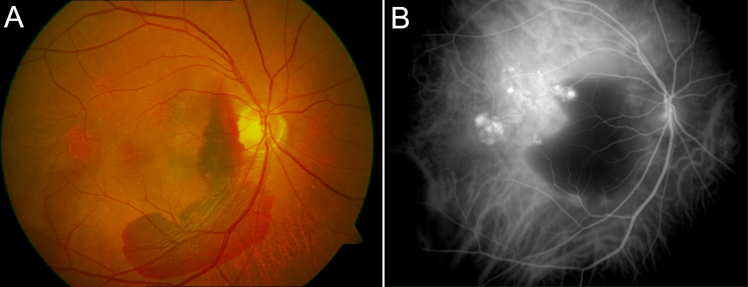
 Figure 1 of
Li, Mol Vis 2010; 16:231-239.
Figure 1 of
Li, Mol Vis 2010; 16:231-239.