Figure 7. Without lens, endogenous Wnt2b expression is lost from anterior optic cup lip. A: A Whole mount view of the eye 24 h after lens removal. B: A section through eye in A showing loss of endogenous Wnt2b gene expression from lip (red-) and cornea (white-arrowheads). Signal is still retained
in non-cornea surface ectoderm (red asterisk). C: An adjacent section showing collagen IX gene expression in lip (red arrowhead). Black arrow indicates a collagen IX positive
piece of lip from a tissue fold, caught on section. D: Whole mount view 48 h after surface ectoderm removal. E: Section through the eye in D, showing retention of Wnt2b signal in lip (red arrowhead). The endogenous signal is lost from lens ectoderm (arrows). F: An adjacent section, shows collagen IX staining (red arrowhead). G-H: Adjacent section analysis of the post surgical anterior optic cup lip, 48 h after lens removal, as in A. G: A bright-field view of the post-surgical optic cup lip, shows that there is no lens. Dashes are used to outline the extent
of tissue neuroepithellial tissue. The outer layer has endogenous pigmentation typical of the retinal pigment epithelium (RPE).
This inner layer is contiguous with the neural retina. H: CollagenIX expression on an adjacent section (red arrowhead) is found in the outer layer of the optic cup lip after lens
removal. The white asterisk marks non-signal. I: A section stained for Wnt2b expression, shows that Wnt2b is not found in the lens extirpated optic cup lip, even when overstained
(white arrowhead). J: An adjacent section immunostained for the RPE marker NaK-ATPase (red arrowheads, see L) shows that the outer layer correctly expresses the RPE marker. K: The expression of the ciliary body marker connexin43. Cx43 is highly expressed in only in the outer layer of the anterior
optic cup (red arrow) and is aberrantly absent from the inner layer (white arrowhead; see M). L: NaK-ATPase expression in control optic cup lip. Expression (Red signal) is found in RPE layer. M: Connexin43 expression in control optic cup lip. Expression (white signal) is found in a line between outer RPE layer (o)
and inner ciliary body layer (i). L denotes lens. Inset box shows that the posterior neural retina does not express detectable
levels of Cx43. Scale bars in B and E are equal to 200 µm, and in G, L and M are equal to 100 µm.
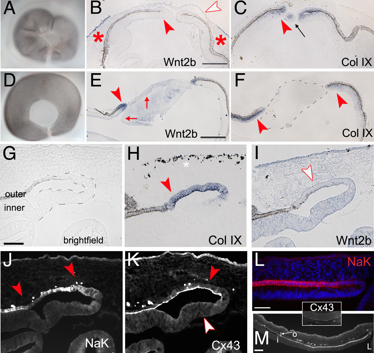
 Figure 7 of
Kitamoto, Mol Vis 2010; 16:2701-2717.
Figure 7 of
Kitamoto, Mol Vis 2010; 16:2701-2717.