Figure 6. Wnt2b is preferentially maintained in anterior transition zones. A: An infected eye at Hamburger-Hamilton (HH) stage 25 with a virus associated depigmented patch. B: The same eye as in A, after in situ hybridization to localize Wnt2b expression. The white asterisks indicate endogenous Wnt2b expression in unaffected
anterior optic cup lip. The boxed area is magnified in C. C: Only the transition zone closest to the lens expresses ectopic Wnt2b (green arrowhead). D,E: An infected eye at HH stage 25, before (D) and after (E) in situ hybridization to localize collagen IX expression. The white asterisks indicate endogenous collagen IX expression
in unaffected anterior optic cup lip tissue. The boxed area is magnified in F. F: CollagenIX is ectopically expressed throughout the borders of the infected patch (green arrows). G, H: An infected eye at HH stage 23 before (G) and after (H) in situ hybridization to localize Wnt2b expression. The asterisks indicate endogenous expression, and the boxed area is
magnified in I. I: At HH stage 23, Wnt2b is ectopically expressed throughout a large portion of the border area, with posterior borders less
robust (white arrowhead) and anterior borders more robust (green arrowhead). A green asterisk indicates native expression
in retinal pigment epithelium (RPE). Red asterisk indicates where RPE is no longer expressing Wnt2b. Transition zones in this
area (red arrow) express Wnt2b. The black arrow indicates a depigmented patch at the lip that does not express Wnt2b. Scale
bars in all panels are equal to 500 µm.
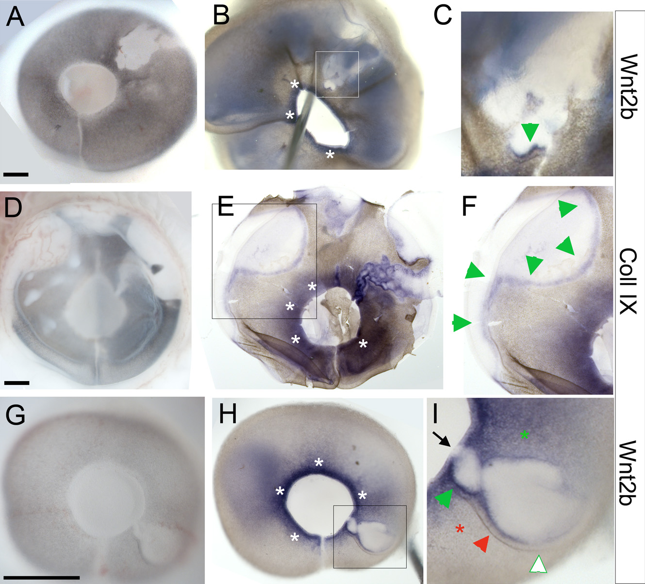
 Figure 6 of
Kitamoto, Mol Vis 2010; 16:2701-2717.
Figure 6 of
Kitamoto, Mol Vis 2010; 16:2701-2717.