Figure 4. Wnt2b expression is not dependent on presence of lens tissue. A: An example of the pre-lens ectoderm removal surgery with red dots indicating the extent of ectoderm removal. B: The operated eye 48 h after re-incubation. C: The contralateral control eye and D: lens-less operated eye stained for Wnt2b expression 24 h after surgery. Note that in these sections the eye flattened and
the vitreous space is not obvious. Expression is found in the outer layer of the anterior optic cup (single arrowheads) and
is more robust in the optic cup lip in the control eye (double arrowheads). An asterisk indicates non-lens tissue. E: Contralateral control and F: lens-less operated eye, 48 h after surgery. The lens-less eye is smaller and the neural retina has formed folds that occupy
the center (dashed lines). Wnt2b gene expression is present at the junction of pigmented (green asterisk) and non-pigmented
(red asterisk) tissue but reduced (overstained section). A typical-looking optic cup lip, with normal hinge formation and
Wnt2b expression is indicated by the single arrowhead and double arrowheads indicate expression without hinge formation. In
E, arrow indicates an artifact rip in the section. G: Contralateral control and H lens-less eye of embryo stained for collagen IX gene expression. G and H are sections adjacent to E and F. Expression is found in same domains where Wnt2b was seen. Scale bars in A and B are equal to 250 µm, and in C, E, F are equal to 200 µm.
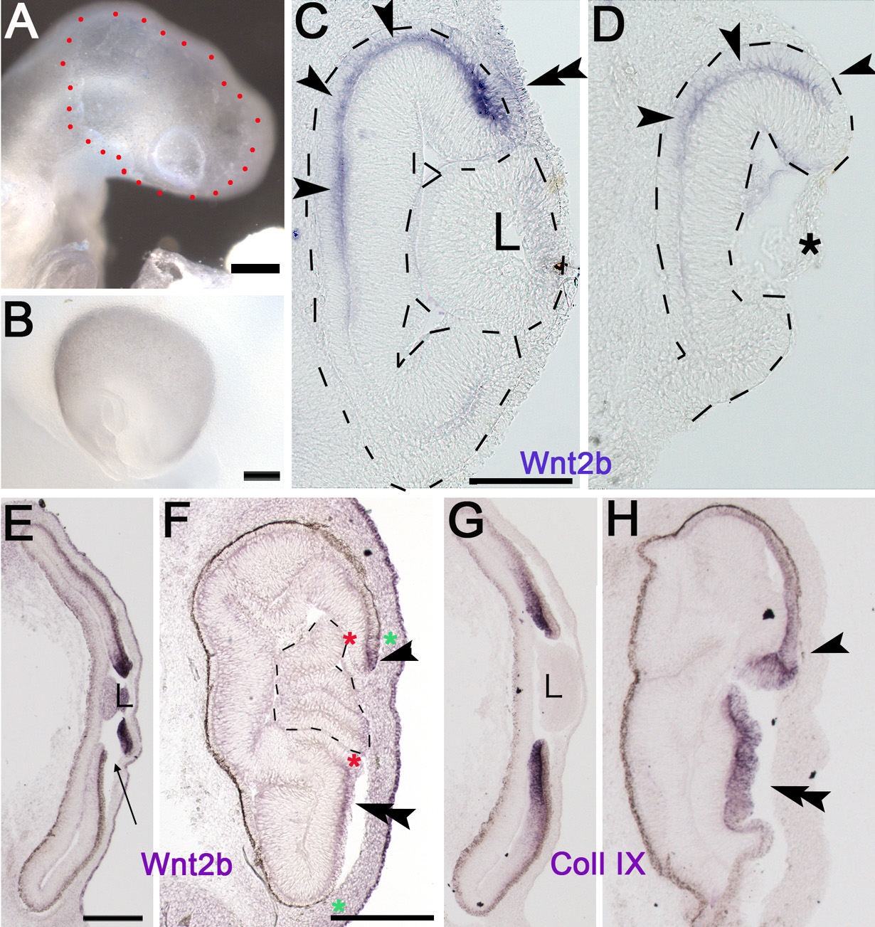
 Figure 4 of
Kitamoto, Mol Vis 2010; 16:2701-2717.
Figure 4 of
Kitamoto, Mol Vis 2010; 16:2701-2717.