Figure 3. The lens cannot induce Wnt2b expression in anterior optic cup lip. A: A highly infected eye with a large patch such that the entire nasal hemisphere (green arrows) is depigmented. B: A second highly infected eye after Xgal staining. C: Similarly infected eye at Hamburger-Hamilton (HH) stage 25, unstained and D: after in situ hybridization to localize collagen IX expression. A red dot and dash orient between the two panels. CollagenIX
expression is found in the depigmented optic cup lip (green arrows in D). White asterisks indicate collagen IX expression in the unaffected anterior, which serves as a native internal control.
E: A highly infected eye at HH stage 25, unstained and F: after in situ hybridization to localize Wnt2b expression. A red dot and dash orient between the two panels, white asterisks
indicate native Wnt2b expression in the unaffected anterior cup lip and green arrows indicate lack of Wnt2b expression in
the depigmented lip. The section plane position for panels G-I are indicated. G: A section through unaffected optic cup lip shows endogenous expression of Wnt2b (arrow) at the hinge between outer retinal
pigment epithelium (RPE) layer and inner ciliary body tissue. H,I: A section through the mis-specified optic cup lip, showing no Wnt2b expression in the hinge (double arrowheads). The outer
layer is completely depigmented and dots demarcate the border between the two epithelia. Scale bars are equal to 50 µm.
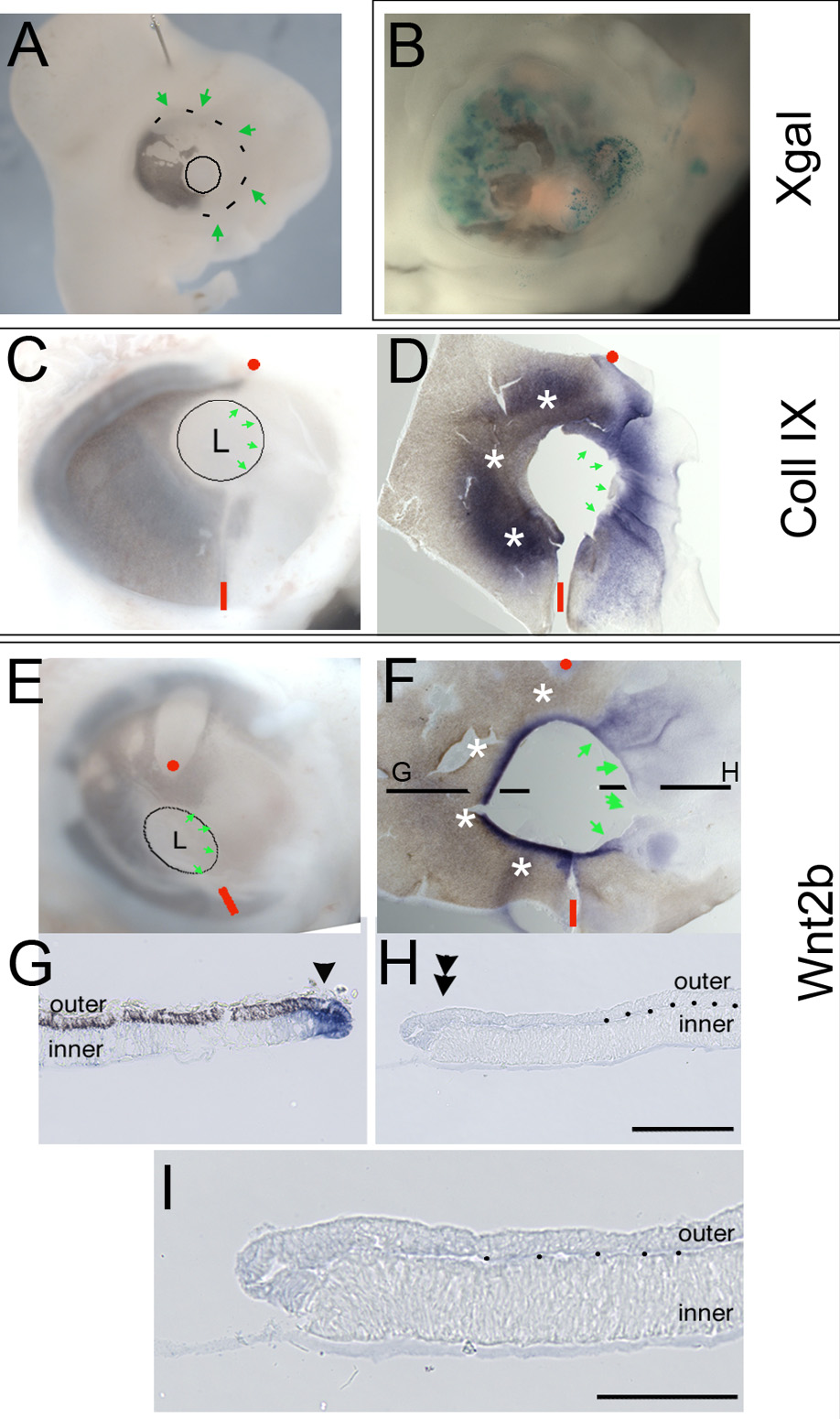
 Figure 3 of
Kitamoto, Mol Vis 2010; 16:2701-2717.
Figure 3 of
Kitamoto, Mol Vis 2010; 16:2701-2717.