Figure 3. Synaptic ribbon proteins
co-distribute with RIBEYE. A, B: Representative images
of the outer plexiform layer (OPL) with the photoreceptor ribbon
synapses triple labeled for RIBEYE (green), Piccolo (red), and Bassoon
(blue) from untreated (A) and EGTA-treated (B) retinae.
Under EGTA treatment, both RIBEYE and Piccolo change from a
horseshoe-shaped to a punctate appearance, even when a Bassoon
horseshoe shape is still visible (B). C-H:
Representative images of the OPL with the photoreceptor ribbon synapses
double labeled for RIBEYE (green) and ribbon associated proteins (red)
from untreated (C, E, G) and EGTA-treated (D,
F, H) retinae. Like RIBEYE and Piccolo, RIM1, CtBP1, and
KIF3A also change from a horseshoe-shaped to a punctate appearance
under EGTA treatment and co-localize with the RIBEYE aggregates. Scale
bar: 5 µm.
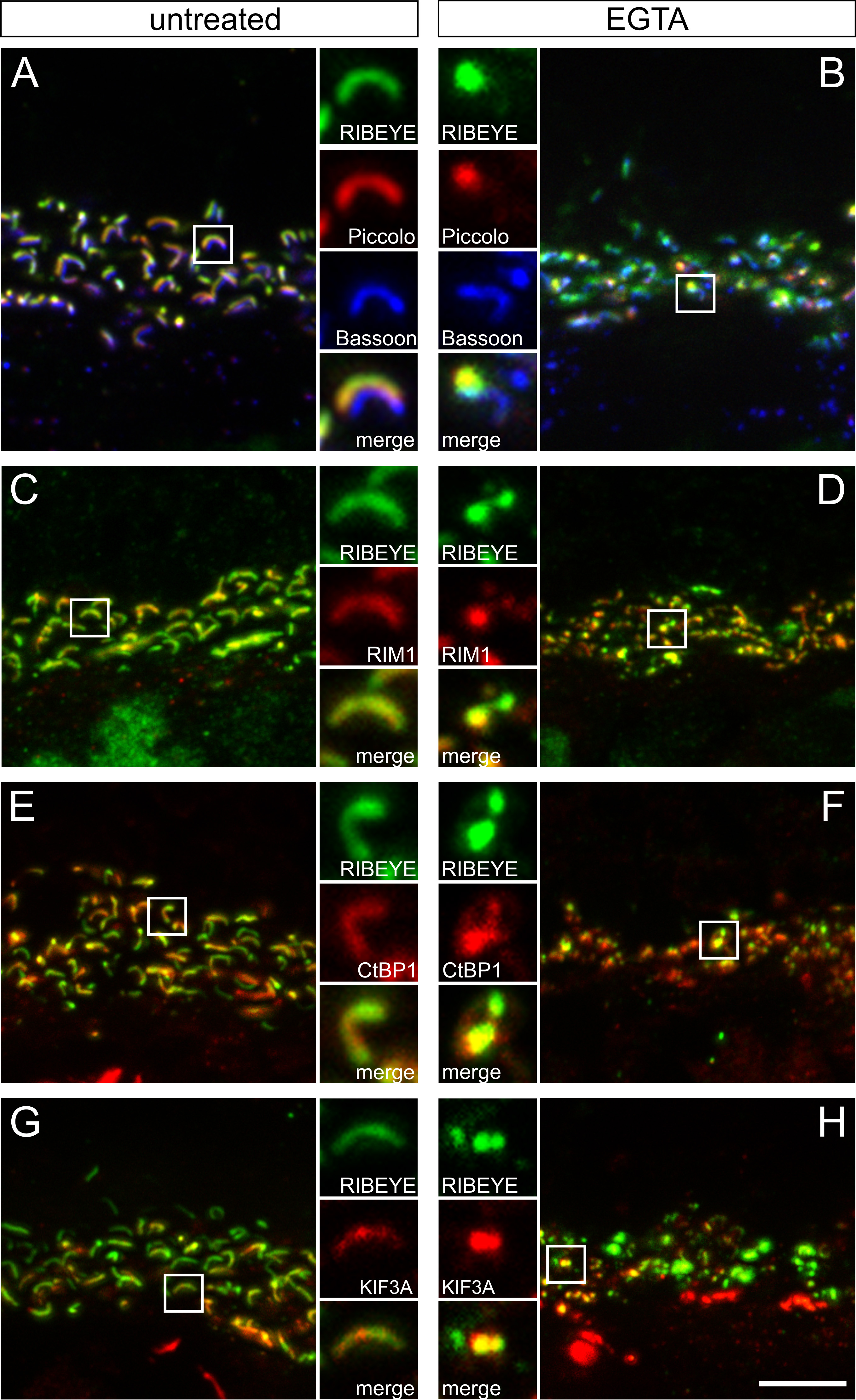
 Figure 3 of Regus-Leidig, Mol Vis 2010; 16:2690-2700.