Figure 2. Bassoon localization at the rod
photoreceptor ribbon complex is more stable than RIBEYE localization. A,
B: Representative images of the outer plexiform layer (OPL) with
the photoreceptor ribbon synapses double labeled for RIBEYE (magenta)
and Bassoon (green) from untreated (A) and EGTA-treated (B)
retinae.
Horseshoe-shaped RIBEYE lines the horseshoe-shaped Bassoon in
untreated retina (A). In EGTA-treated retina, RIBEYE often
disintegrates into several aggregates (B). C, D:
Immunoelectron micrographs of rod photoreceptor ribbon synapses from
EGTA-treated retina. RIBEYE labeling (C) is found at the
spherical material pinching off the ribbon (arrowhead) as well as at
the active zone (arrows). In contrast, Bassoon labeling (D) is
concentrated at the ribbon base (arrows) and the material pinching off
the ribbon is not stained for Bassoon (arrowhead). E:
Quantification of the Bassoon/RIBEYE co-localization in untreated and
EGTA-treated retinae. In double stained vertical sections, either
horseshoe-shaped (HS) RIBEYE or horseshoe-shaped Bassoon profiles were
counted and checked for the morphological appearance of the respective
other marker. When counts were based on horseshoe-shaped Bassoon (bar
groups 1 to 3), the fraction of sites where RIBEYE staining appears in
a horseshoe shape decreases under EGTA conditions, whereas the punctate
(club-shaped, CS, or spherical-shaped, SS) and cloudy appearance of
RIBEYE staining increases concomitantly. When counts were based on
horseshoe-shaped RIBEYE (bar group 4), only a negligible fraction of
sites had no horseshoe-shaped Bassoon under both conditions. The total
number of horseshoe-shaped Bassoon sites in EGTA-treated retinae was
reduced in the same OPL volume. F, G: Representative
images of the OPL double labeled for the rod photoreceptor terminal
marker Veli3 (magenta) and Bassoon (green) from untreated (F)
and EGTA-treated (G) retinae. In the EGTA-treated retina, Veli3
labeled rod photoreceptor terminals often show punctate or a lack of
Bassoon labeling, which is in contrast to the horseshoe-shaped Bassoon
labeling present in rod photoreceptor terminals of untreated retina.
Scale bars: 5 µm (A, B, F, G), 0.2 µm (C,
D).
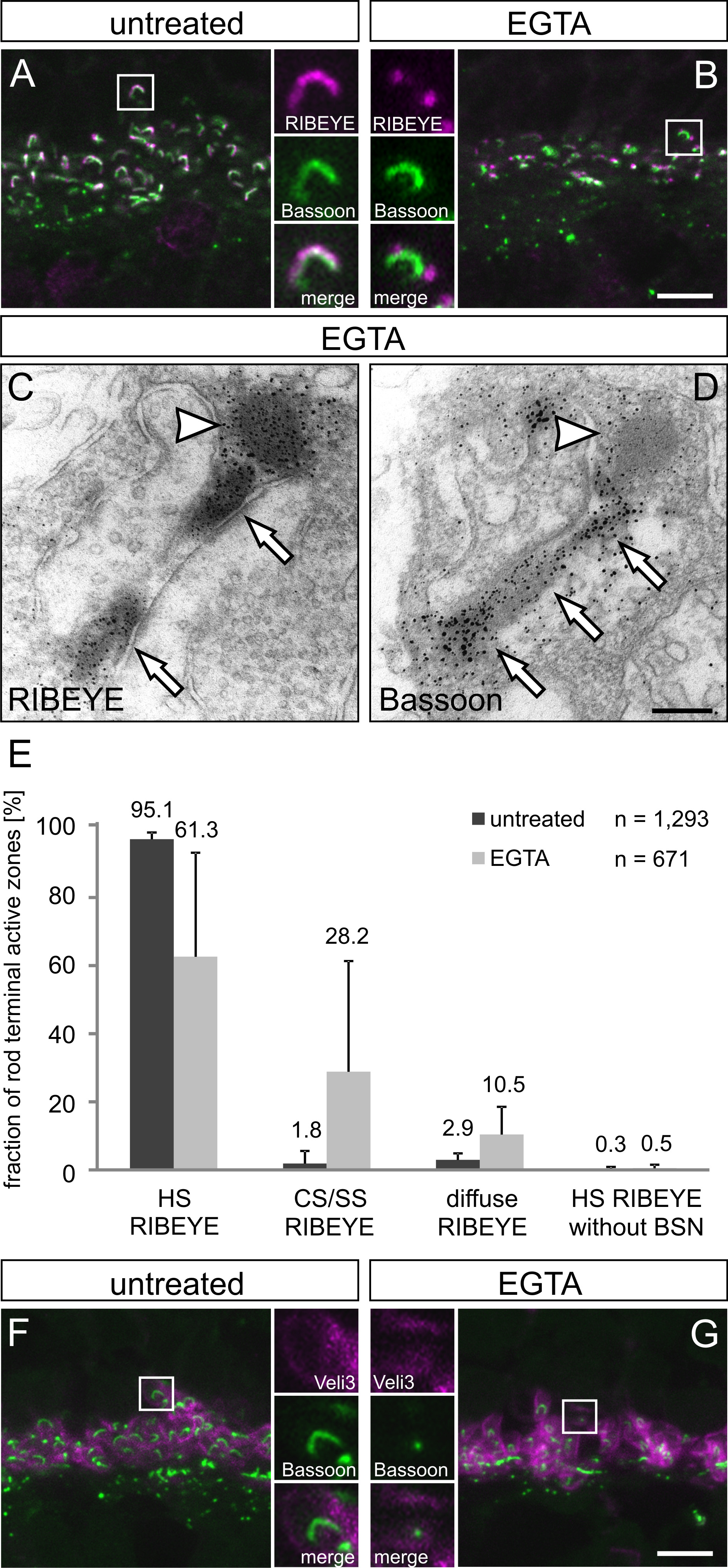
 Figure 2 of Regus-Leidig, Mol Vis 2010; 16:2690-2700.