Figure 1. Enrichment of club-shaped and
spherical-shaped ribbon material using EGTA treatment. A: The
percentage of rod-shaped ribbons is significantly reduced in
EGTA-treated retinas (EGTA) compared to ionophore A23187-treated or
untreated retinas, while the percentage of club-shaped and
spherical-shaped ribbon profiles is significantly increased (n=237
ribbon profiles for EGTA- and ionophore-treated retinas; n=294 ribbon
profiles for untreated retinas; error bars=SD). B-G:
Electron micrographs of rod (B, C, D) and cone (E,
F, G) photoreceptor terminals, showing examples of
different ribbon states (arrowheads): rod-shaped (B, E),
club-shaped (C, F), and spherical-shaped (D, G).
*
p<0.05, ** p<0.01, *** p<0.001. Scale bars: 0.2 µm.
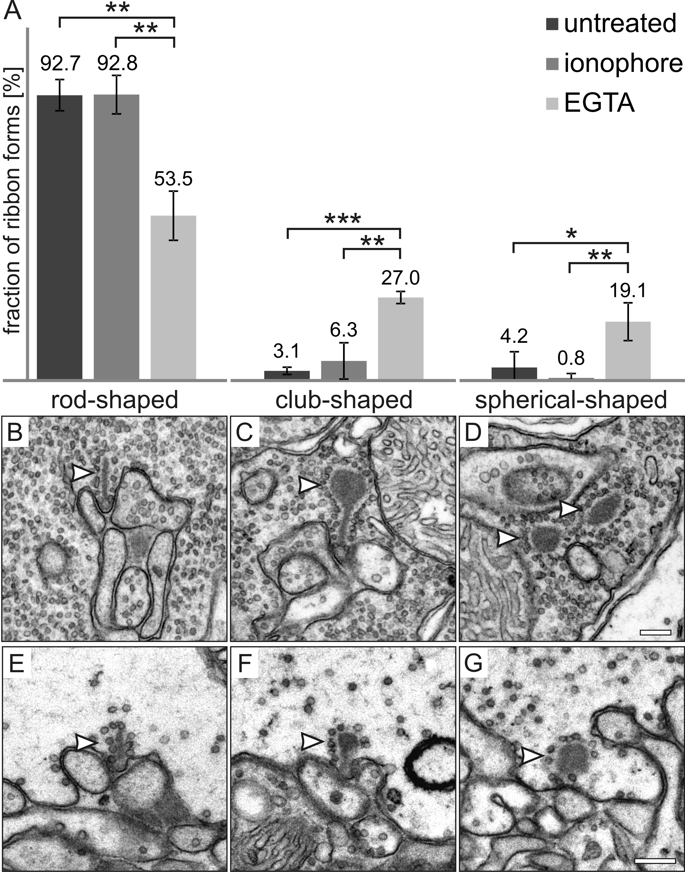
 Figure 1 of Regus-Leidig, Mol Vis 2010; 16:2690-2700.