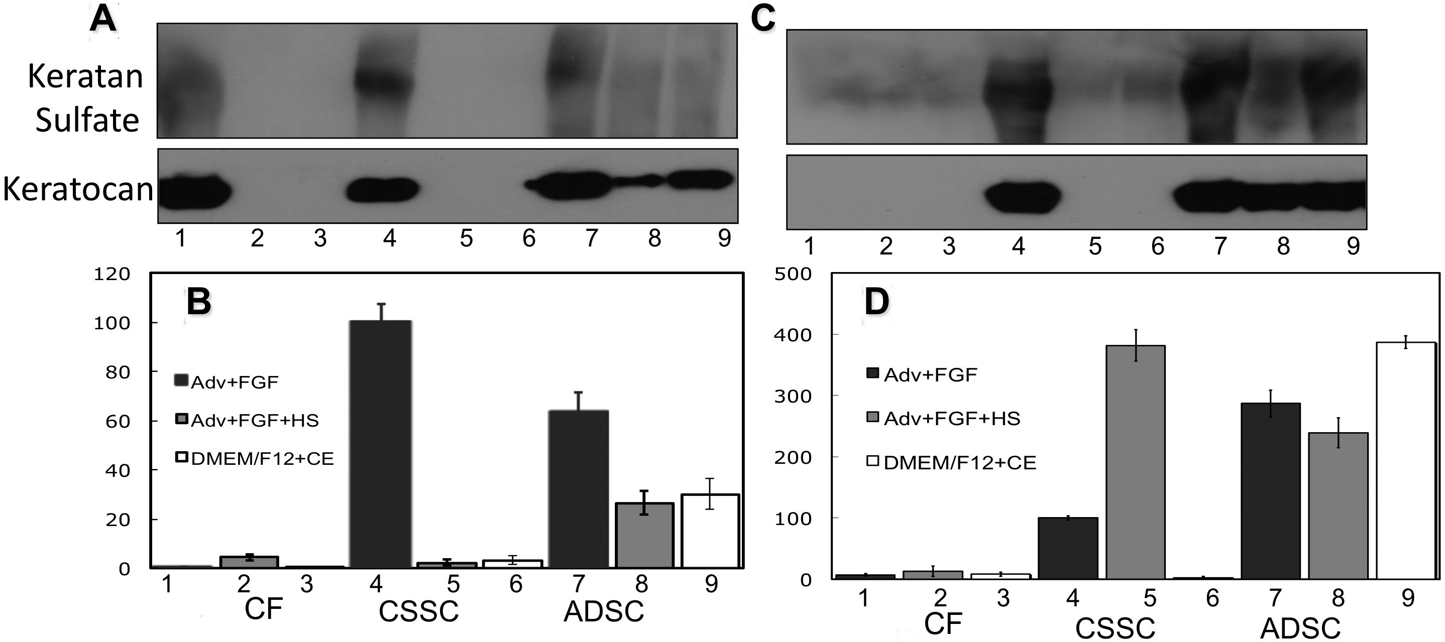
Figure 5. Keratan sulfate, keratocan
protein, and mRNA expression by CSSC, ADSC, and CF in different media.
A,
B: Fibrin gel cultures after 3 weeks.
C,
D:
Pellet cultures after 3 weeks.
A and
C are western
blots showing keratan sulfate and keratocan.
B and
D
show qPCR data of keratocan mRNA. Samples 1–3: CF, 4–6: CSSC cells;
7–9, ADSC cells. Samples 1, 4, 7: keratocyte differentiation medium;
Samples 2, 5, 8: keratocyte differentiation medium + 1% HSHS. Samples
3, 6, 9: DMEM/F-12 medium with bovine corneal extract (1:10) [
25]. Expression of
mRNA is shown normalized to monolayer of CSSC in keratocyte
differentiation medium=100.
 Figure 5 of Du, Mol Vis 2010; 16:2680-2689.