Figure 4. Induction of keratocyte markers
in ADSC. A, B: ADSC were cultured in fibrin gels for 3
weeks in keratocyte differentiation medium. C, D: ADSC
were cultured as pellet for 3 weeks in keratocyte differentiation
medium. Immunofluorescent staining shows the presence of keratan
sulfate with antibody J19 (green; A, C) or keratocan
with antibody KeraC, (green; B, D). Red shows nuclear
staining by propidium iodide. E: RT–PCR shows keratocan
expression in (left to right) uncultured keratocytes (positive
control), ADSC in fibrin gel, ADSC as pellet culture, ADSC in SCGM.
Scale bars=20 μm.
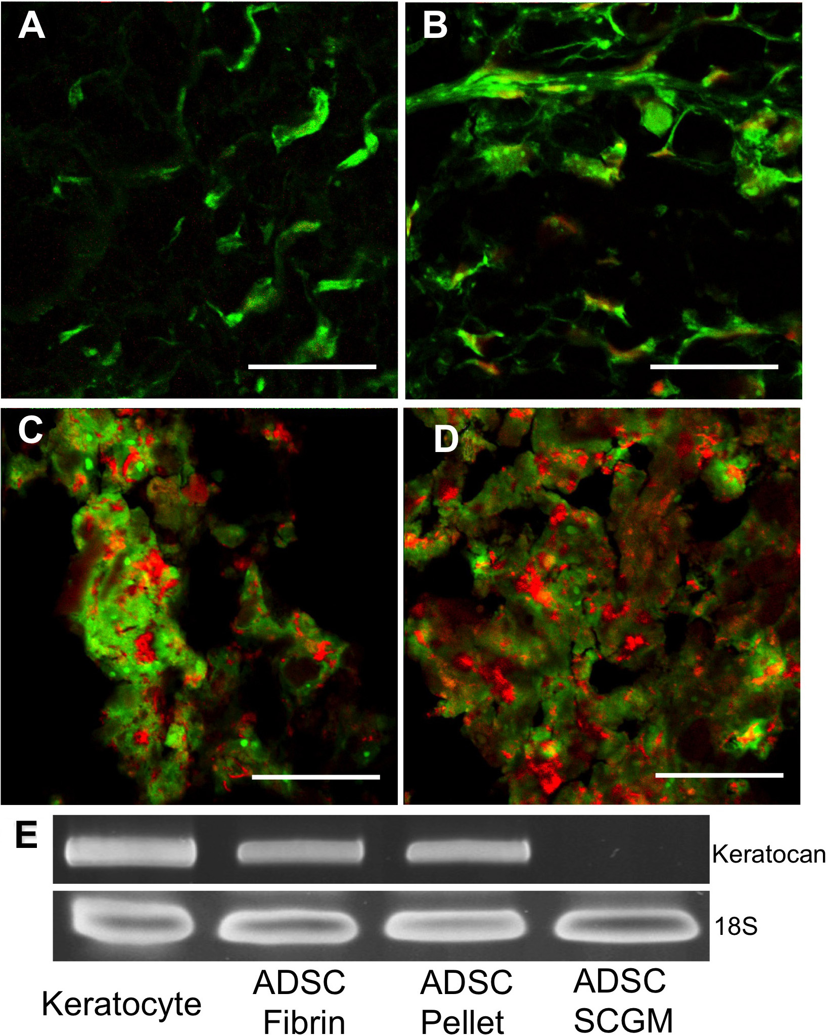
 Figure 4 of Du, Mol Vis 2010; 16:2680-2689.