Figure 3. Induction of cartilage matrix
expression by ADSC. ADSC (A) and CF (B) were cultured as
pellets (2×10⁵) in chondrocyte differentiation medium for
three weeks. The pellets were fixed, imbedded in OCT, cut into 8 µm
thick sections and stained with toluidine blue to detect proteoglycan
staining typical of cartilage. Scale bar indicates 50 μm.
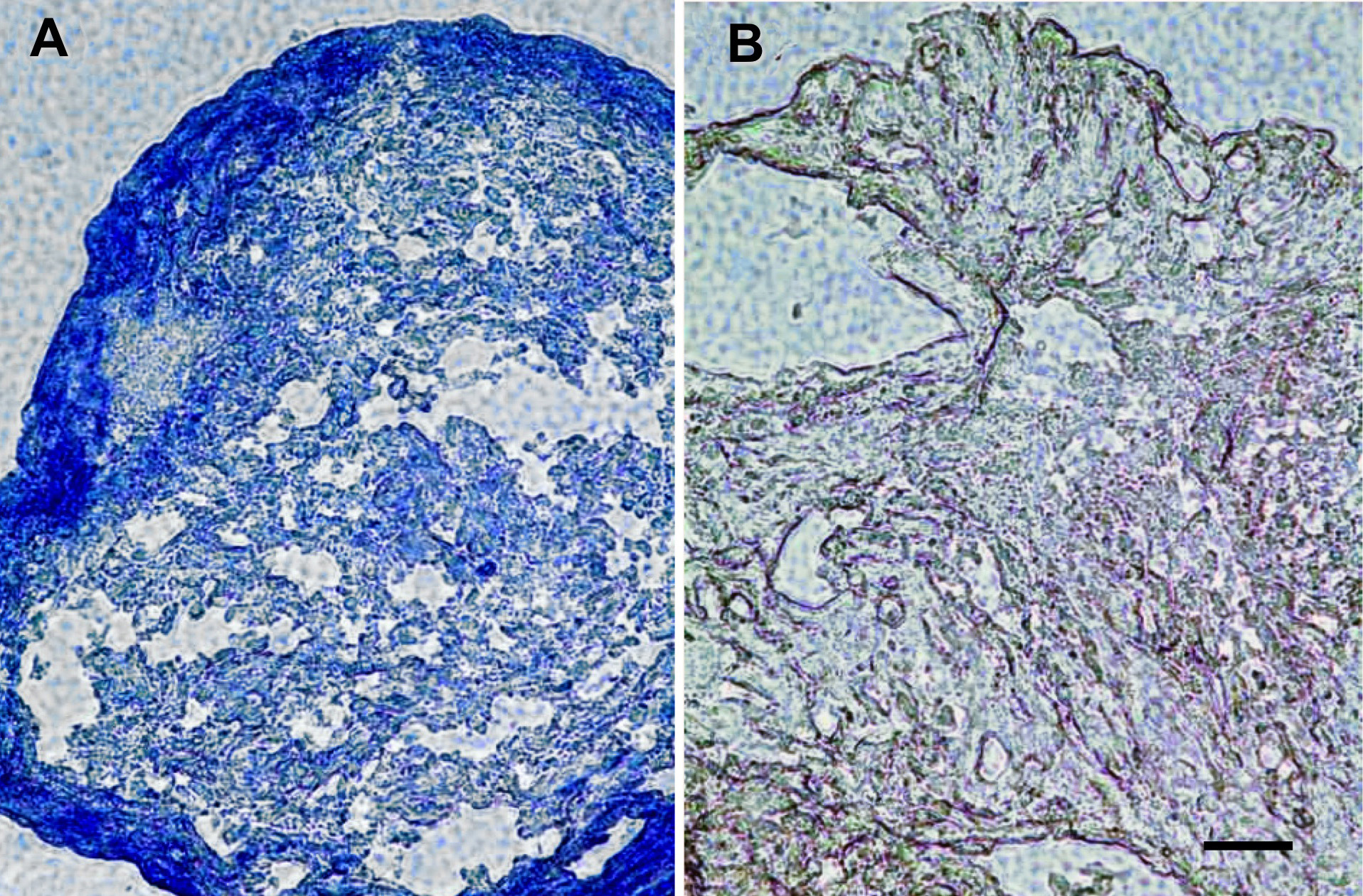
 Figure 3 of Du, Mol Vis 2010; 16:2680-2689.