Figure 2. Immunocytochemistry was used to
illustrate altered expression of VE-cadherin, β-catenin, vimentin,
N-cadherin and protein kinase B (AKT2) in monkey choroid-retinal
endothelial cells (RF/6A) and human umbilical vein endothelial cells
(HUVEC) after the treatment by advanced glycation end products of BSA
(AGE-BSA; 200 mg/l) for 24 h. For RF/6A cells, there were decreased
expression of markers of endothelial cells, VE-cadherin (A) and
β-catenin (B), and increased expression of markers of
mesenchymal cells, N-cadherin (C) and vimentin (D).
Similar to the RF/6A cells, the protein expression of VE-cadherin (F)
and
β-catenin (G) in the HUVEC cells was downregulated, and the
expression of N-cadherin (H) and vimentin (I) in the
HUVEC cells was upregulated. At the same time, the expression of Akt2
in the both cells increased (E, J). Additionally, in the
RF/6A cells, the decreased part of β-catenin was mainly in the
cytoplasm, while the protein expressed in the nucleus changed little (B);
after
treatment with AGE-BSA, N-cadherin expression was upregulated in
the nucleus and downregulated in the intracytoplasm (D). In the
HUVEC cells, there were no such phenomena.
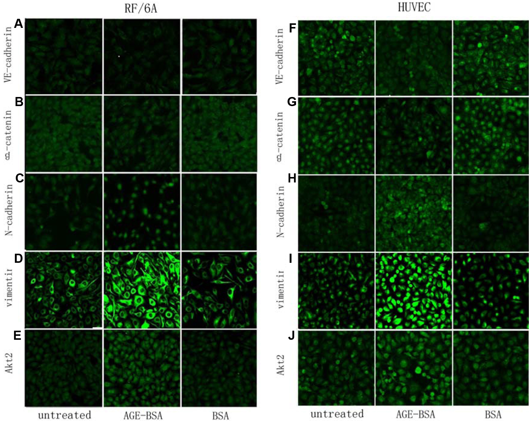
 Figure 2 of Ma, Mol Vis 2010; 16:2669-2679.