Figure 3. Color (left) and
autofluorescence (right) fundus photographs from the right eye of
patient II-6, recorded at the age of 35 years. Fluorescence was absent
in a hyperpigmented central zone of outer retinal atrophy, whereas an
irregular gray to white surrounding zone showed confluent
hyperautofluorescence. The general level of fundus
autofluorescence outside the macula appeared to be normal.
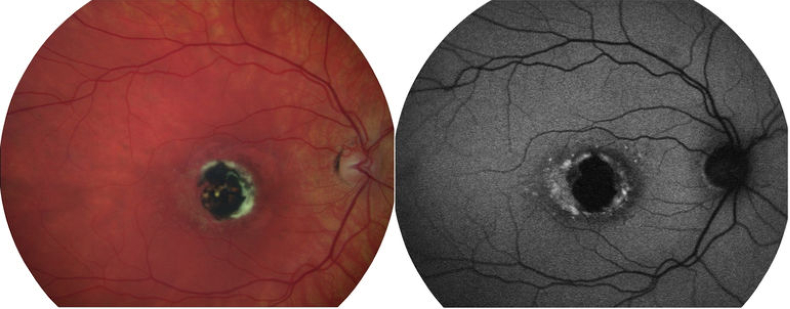
 Figure 3 of Rosenberg, Mol Vis 2010; 16:2659-2668.