Figure 2. Selected color fundus pictures
of the North Carolina macular dystrophy pedigree. Right (left column)
and left (right column) macular regions are shown for four affected
individuals: A-B, patient II-1; C-D,
patient II-6; E-F, patient III-2; G-H,
patient III-3. The macular lesions A-B and E-H
are grade 1, while the lesions in C-D are grade 3.
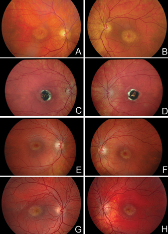
 Figure 2 of Rosenberg, Mol Vis 2010; 16:2659-2668.