Figure 2. The CT scan and MRI scan results. A: CT scan of the proband’s brain; these images show cerebellar atrophy with widened and deepened sulci and permit observation
of the fifth ventricle. Arrows show cerebellar atrophy is characterized by narrowed lobes and widened folds of cerebellar
hemisphere and vermis; and the evolution of the cavity of septum pellucidum. B: MRI scan of carrier III:6’s brain; no abnormality was observed.
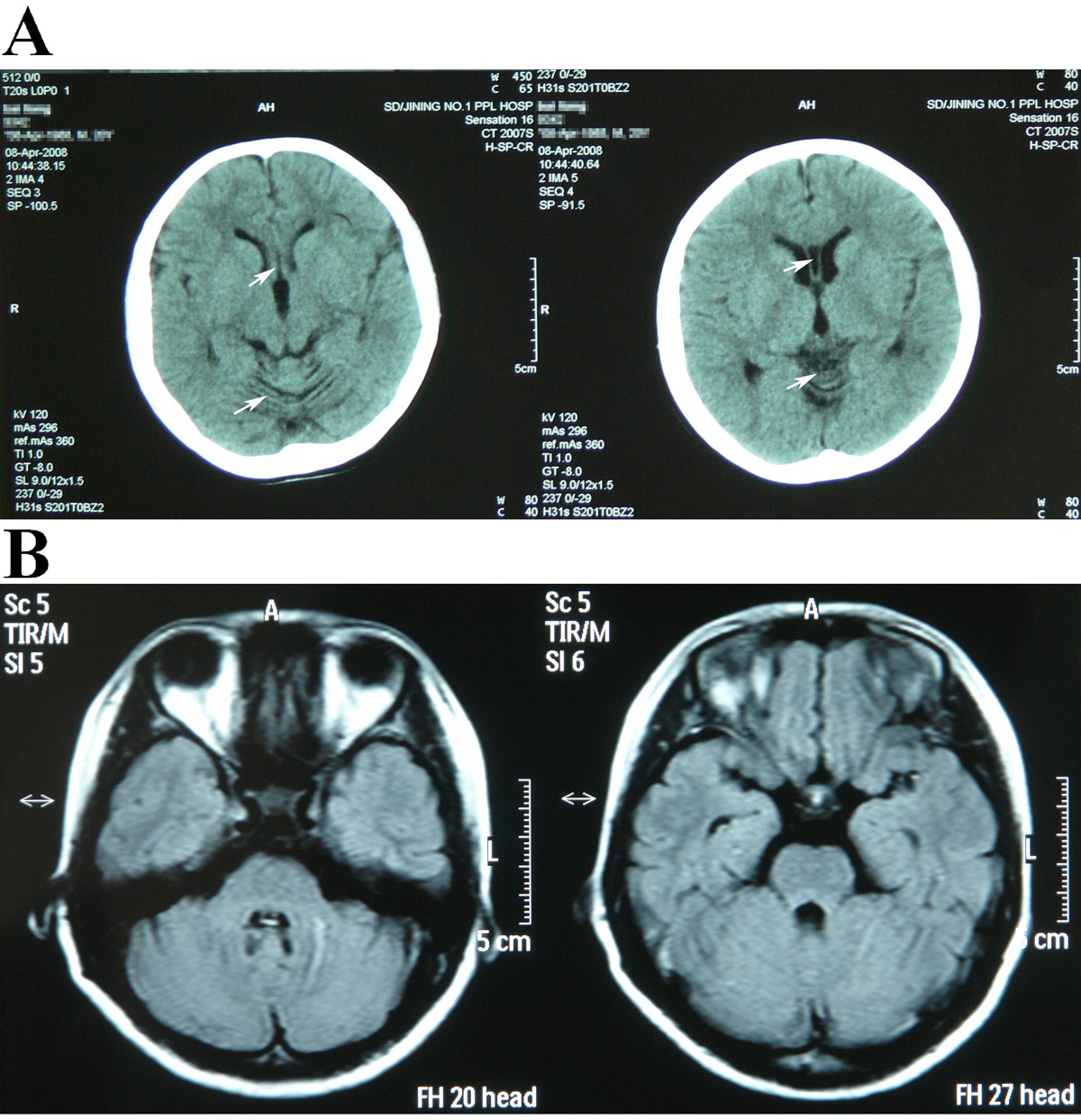
 Figure 2 of
Liu, Mol Vis 2010; 16:2653-2658.
Figure 2 of
Liu, Mol Vis 2010; 16:2653-2658.