Figure 4. Optical coherence tomography
(OCT) scans demonstrate protection of ONL by pyruvate and nicotinamide
against LD. A: Representative ONL images were taken from OCT in
control, 7 days after 4 h light damage in non-dark conditions (4 h LD
(light)), 4 h LD (light) + P, 4 h LD (light) + N, and 4 h LD (light) +
L, where P, N, and L refer to 500 mg/kg i.p. injection before LD and
3x/week of pyruvate, nicotinamide, or lactate, respectively. Bar
represents 50 microns. B: The mean thickness in central
superior and central inferior hemispheres (arrow or bar in A)
of the retina in microns after these exposures is presented ± SEM
(n=6). * indicates difference from control, # indicates difference from
light damage, and $ indicates difference between dark maintained and
non-dark maintained retinas at p<0.05 by one-way ANOVA and a
Bonferroni test.
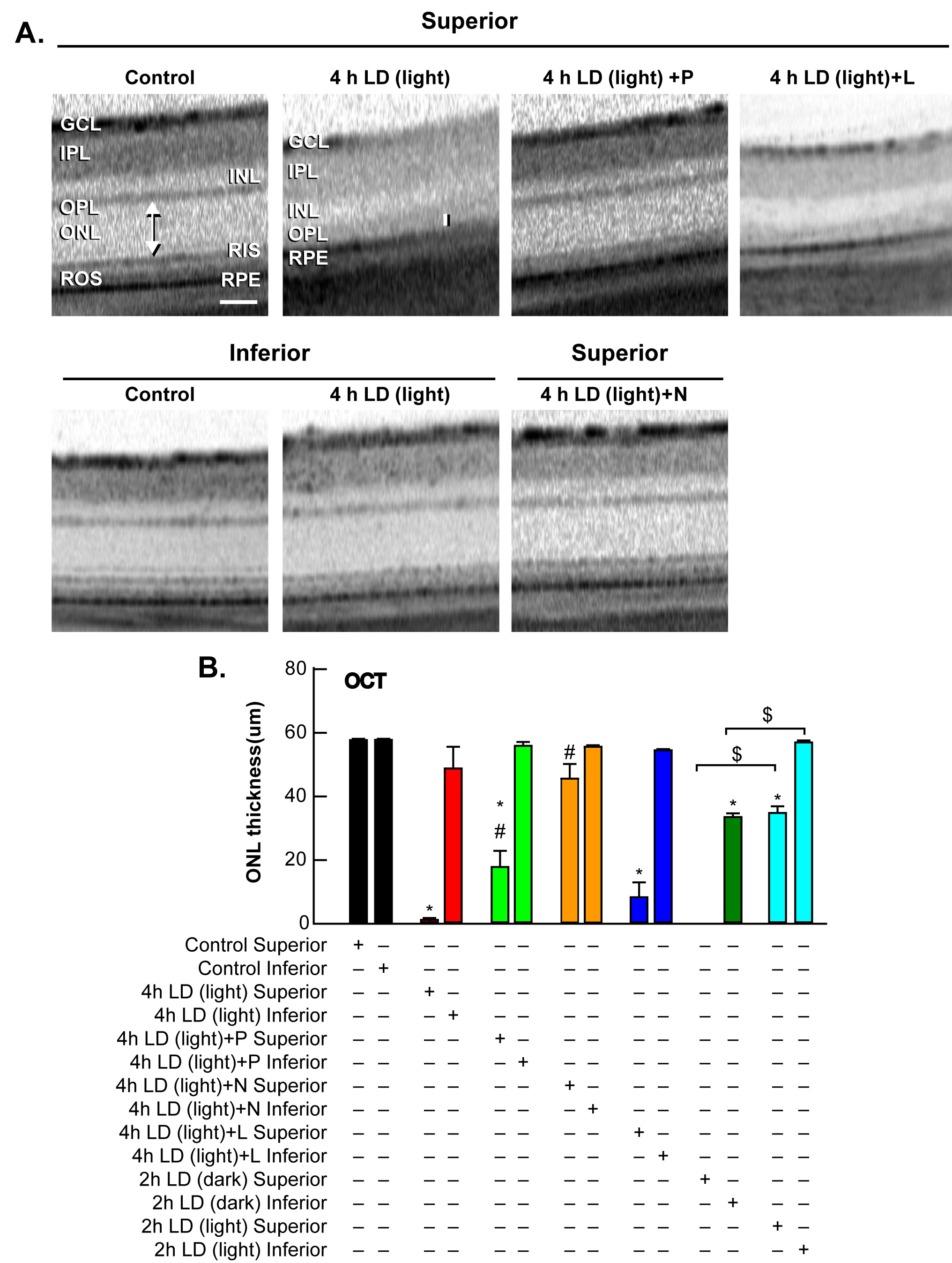
 Figure 4 of Sheline, Mol Vis 2010; 16:2639-2652.