Figure 3. Light-induced damage caused Zn2+
accumulation preferentially in superior photoreceptors, retinal pigment
epithelial (RPE), rod outer segments (ROS), and outer plexiform layer
(OPL). Light-induced damage (LD) was performed and fresh frozen rat
retinas were analyzed at 4 or 14 h after 1 h of light damage in dark
maintained conditions (dark) or after 4 h of light damage in non-dark
maintained conditions (light). Zn2+ accumulation (white
regions) was assessed by ZP1 staining of fresh frozen rat eye cryostat
sections cut at 10 microns, which were dried and stained in 5 μM ZP1
for 2 min. Representative photomicrographs (n=4) were taken of the mid
inferior and mid superior regions of the retina at 5 s exposure for:
control, 4 h LD (light) + 4 h, and 1 h LD (dark) + 4 h; at 1 s exposure
for: 1 h LD (dark) + 14 h; and at 2 s exposure for: 4 h LD (light) + 14
h, and 1 h LD (light) + 14 h. Layers are as marked. Notice the large
increase in the number and intensity of Zn2+ stained cells
in superior ONL (brackets), ROS, and RPE. Light=non-dark maintained,
Dark=dark maintained.
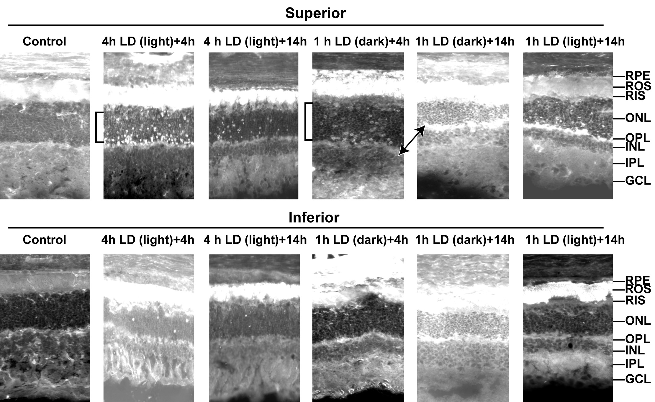
 Figure 3 of Sheline, Mol Vis 2010; 16:2639-2652.