Figure 4. H&E profile of the lens sections. The upper panel (A and B) represents lenses in rats given the galactose diet treated only with the placebo. Apparent is the retention of nucleus in
the fiber cells due to defective cellular transition. The nuclei are also clearly pyknotic. The central area of the tissue
(B) also contains some nucleated cells. Other cells are swollen and detached form each other. These abnormalities were inhibited
in the group treated with caffeine eye drops as shown in the lower panel (C and D).
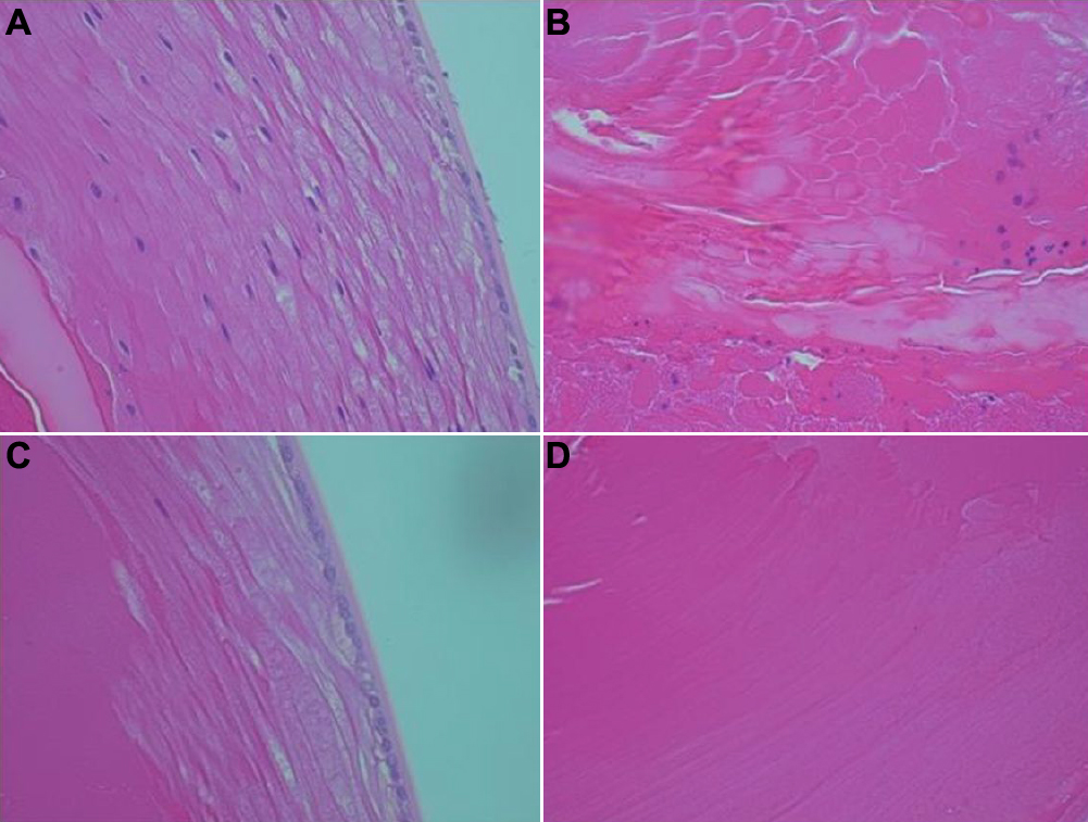
 Figure 4 of
Varma, Mol Vis 2010; 16:2626-2633.
Figure 4 of
Varma, Mol Vis 2010; 16:2626-2633.