Figure 5. Rods and cones immunolabeling in
3-week-old rd10, rd10/wild-type (WT), rd10/human transferrin (hTf), and
TghTf mice. Cones (A-D) and rods (E-H) were
localized on frozen sections from 3-week-old rd10 (A, E),
rd10/WT
(B, F), rd10/hTf (C, G), and
TghTf (D, H) mouse retinas. Cones were labeled with
peanut-agglutinin coupled with fluorescein isothiocyanate and rods with
an antibody against opsin (Rho4D2). Abbreviations: IS is inner segment;
ONL is outer nuclear layer. Scale bars in A-D equal 10
μm; E-H scale bars equal 50 μm.
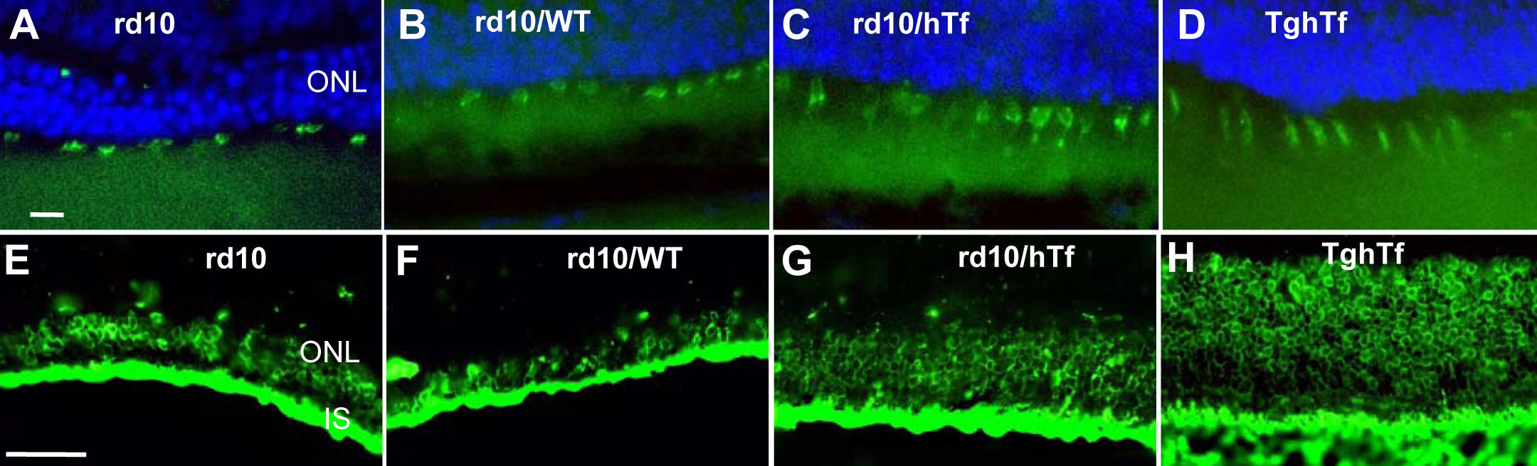
 Figure 5 of Picard, Mol Vis 2010; 16:2612-2625.