Figure 4. Clinical photographs of corneas
in one affected member (I-1) in Family 2 at 63 years of age. The right
eye (A) contained opacifications from presumed recurrent disease
20 years after a penetrating keratoplasty. The left eye (B)
showed a corneal implant that was transparent after a penetrating
keratoplasty 10 years ago. This eye compounded with the primary
cataract and the corrected visual acuity with pinhole is 20/32.
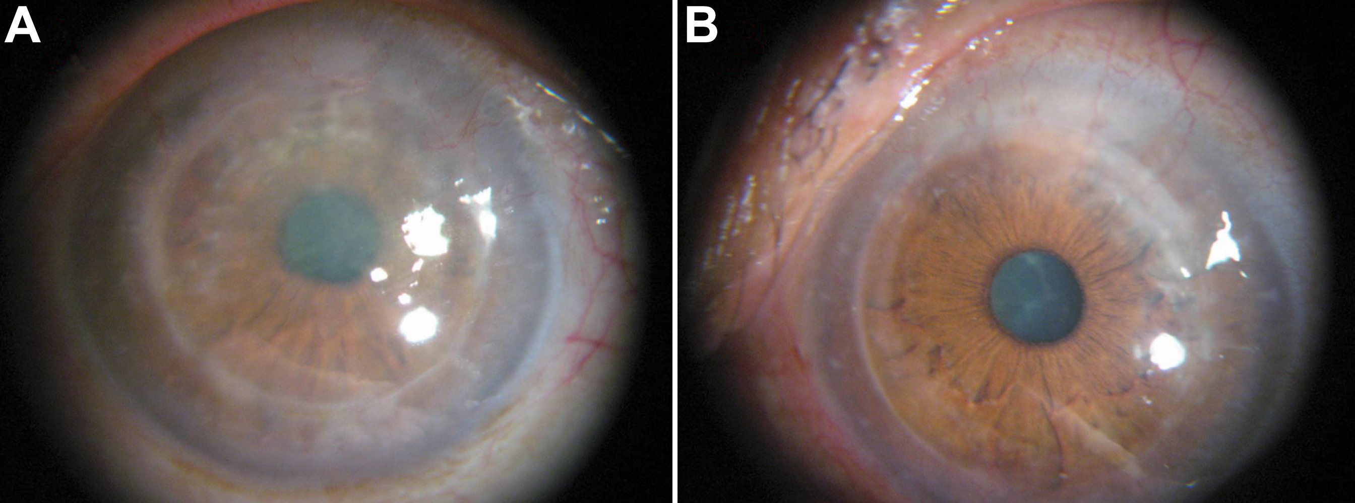
 Figure 4 of Zhong, Mol Vis 2010; 16:224-230.