Figure 6. CERKL expression pattern in the
mouse retina. Serial sagittal sections of adult mouse retina were
immunostained with the RA anti-CERKL antibody. A-C:
CERKL staining (green) shows expression in the photoreceptor cell layer
(PRL), the inner nuclear layer (INL), and the ganglion cell layer
(GCL). D-F: Double staining for CERKL (green) and PAX6
(red), a marker for amacrine cells, in the INL and GCL. G-I:
Double
staining for CERKL (green) and OPN1 (red), a marker for cone
photoreceptor cells. The insert in panel I shows a higher
magnification of a double-stained cone photoreceptor. J-L:
Double
staining for CERKL (green) and RHO (rhodopsin; red), a marker
for rod photoreceptor cells. Nuclei are stained with TO-PRO-3 (blue).
Note that CERKL is highly expressed in cone photoreceptors, while its
expression level in rod photoreceptors is very low. No staining was
observed when sections were stained with serum from pre-immune rabbits
or with secondary antibody only (data not shown). PRL, photoreceptor
layer; ONL, outer nuclear layer; INL, inner nuclear layer; GCL,
ganglion cell layer. Scale bars, 20 µm.
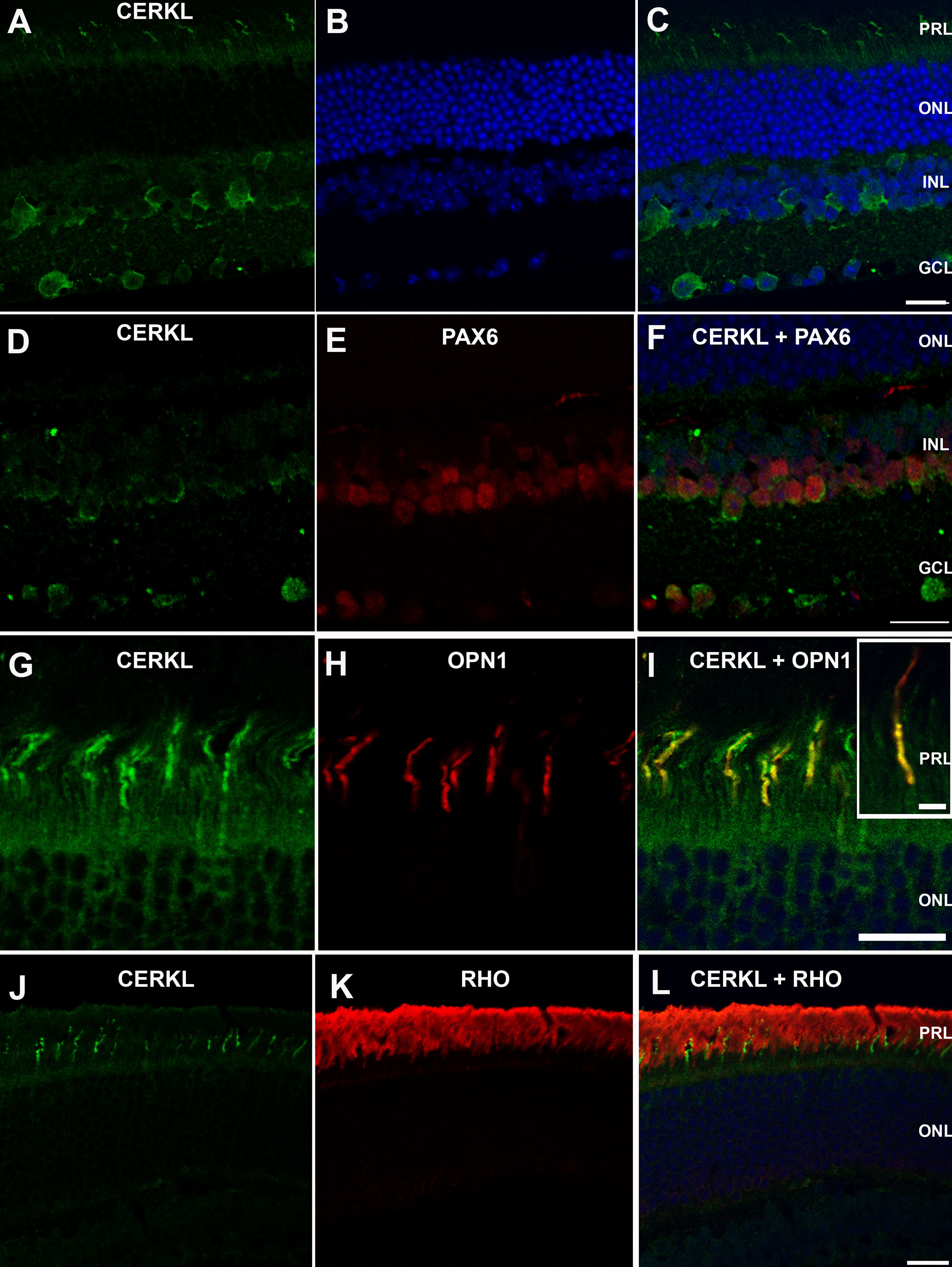
 Figure 6 of Vekslin, Mol Vis 2010; 16:2539-2549.