Figure 5. CERKL subcellular localization
in 661W cells. Cells were stained with the RA anti-CERKL antibody (red)
and with TO-PRO-3 for nuclear staining (blue). A-C:
CERKL can be located in both the nucleus and the cytoplasm, where it is
concentrated in the perinuclear region. Scale bar, 20 µm. D-F:
CERKL
is absent from nucleoli (arrows), which are stained by the
nucleolar marker Fibrillarin (green). Scale bar, 20 µm.
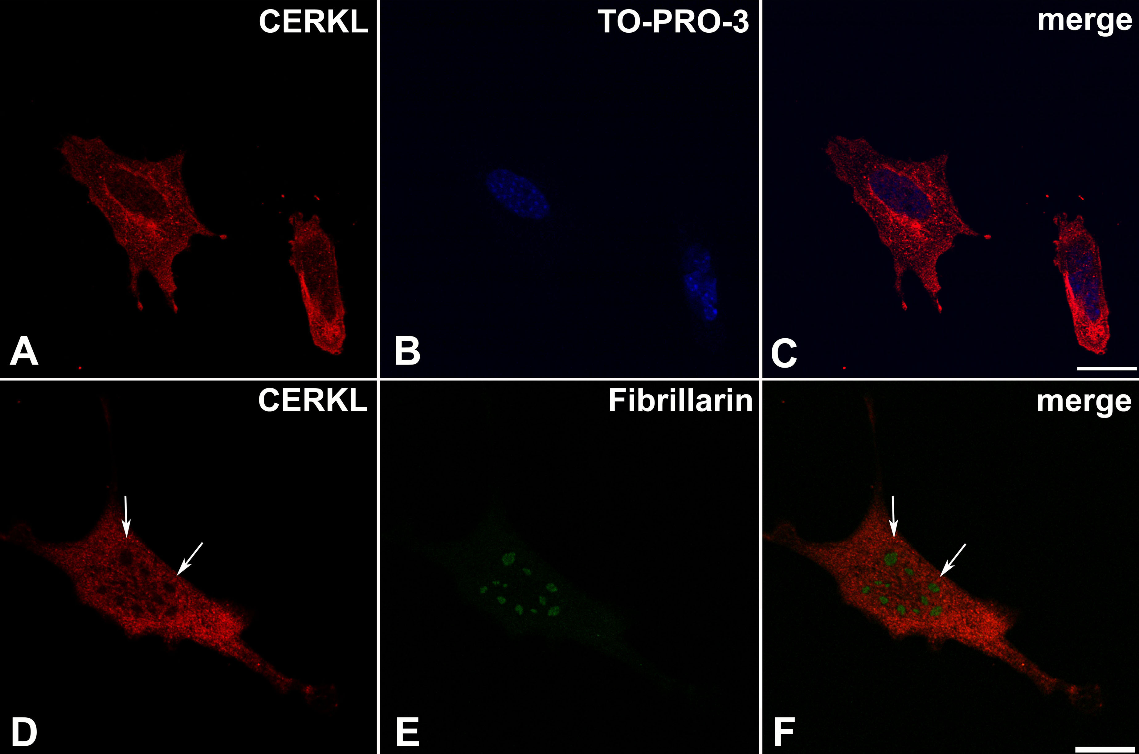
 Figure 5 of Vekslin, Mol Vis 2010; 16:2539-2549.