Figure 4. CERKL subcellular localization
in ARPE-19 cells. Cells were stained with the RA anti-CERKL antibody
(red) and with DAPI for nuclear staining (blue). A-C:
The subcellular localization of CERKL is variable. It can be located in
both the nucleus and the cytoplasm (arrowhead), or only in the
cytoplasm (arrows). In many cells, CERKL is concentrated in the
perinuclear region (asterisks). Scale bar, 50 µm. D-F: A
cell in which CERKL is absent from the nucleus. Scale bar, 10 µm. G-I:
A
cell in which CERKL is concentrated in the nucleus. Scale bar, 10 µm.
J-L: CERKL is absent from nucleoli (arrows), which are
stained by the nucleolar marker Fibrillarin (green). Scale bar, 10 µm.
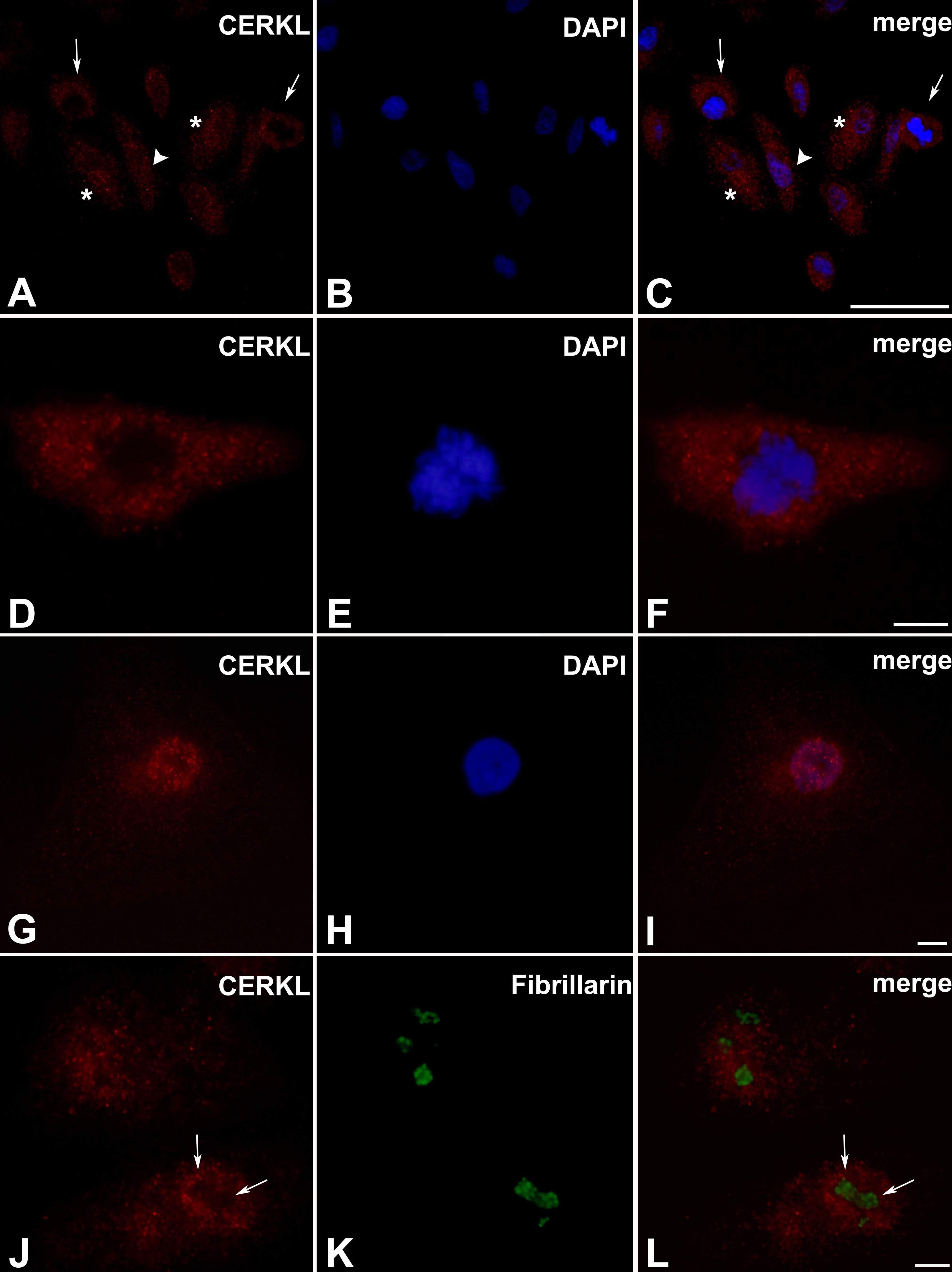
 Figure 4 of Vekslin, Mol Vis 2010; 16:2539-2549.