Figure 5. Spatiotemporal characteristics
of Nε-(3-formyl-3,4-dehydropiperidino)lysine (FDP-lysine) accumulation
in diabetic Müller cells. A and B: Representative
confocal images of FDP-lysine immunoreactivity in retinal sections from
non-diabetic rats 1 and 4 months post injection of citrate buffer. C-F:
Immunohistochemisty
for FDP-lysine in retinal sections from
STZ-diabetic rats 1, 2, 3, and 4 months after the induction of
diabetes. The intensity of FDP-lysine staining initially increased in
the end feet of Müller cells at the ILM after 2 months of diabetes (D,
arrows)
and subsequently spread throughout their inner processes
extending from the ILM to the INL (E, F, arrows). G:
Summary
data revealed that FDP-lysine immunoreactivity was
significantly increased in diabetic retina in regions of the ILM–GCL
and IPL, 2 and 3 months following the onset of diabetes, respectively.
There was no significant difference in FDP-lysine in the outer retina
at any time point between the nondiabetic and diabetic groups. The
scale bars refer to 50 μm.
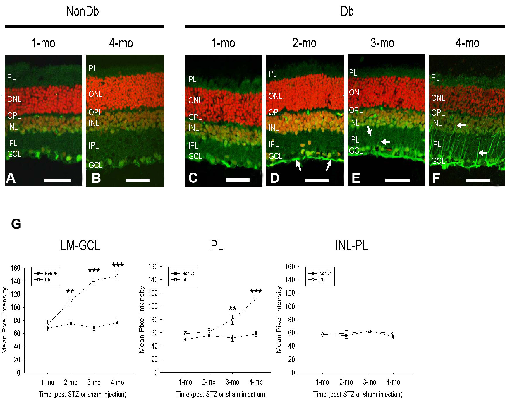
 Figure 5 of Yong, Mol Vis 2010; 16:2524-2538.