Figure 3. Nε-(3-formyl-3,4-dehydropiperidino)lysine (FDP-lysine) immunoreactivity (green fluorescence) in vertical sections of retina
from nondiabetic (NonDb) and diabetic (Db) rats of 4 months disease duration. Nuclei were counterstained with propidium iodide
(red fluorescence). A: Low and high magnification images of FDP-lysine immunoreactivity in the normal rat retina. Strong immunolabelling was detected
in the retinal ganglion cell layer (GCL) and nuclei in the inner nuclear layer (INL). B: Low and high power confocal micrographs of FDP-lysine immunoreactivity in diabetic rat retina. Prominent immunolabelling
appeared at the ILM and in radial processes in the inner retina (arrows). C: Summary data showing that diabetes caused a statistically significant increase in FDP-lysine immunolabelling limited the
innermost retinal layers.
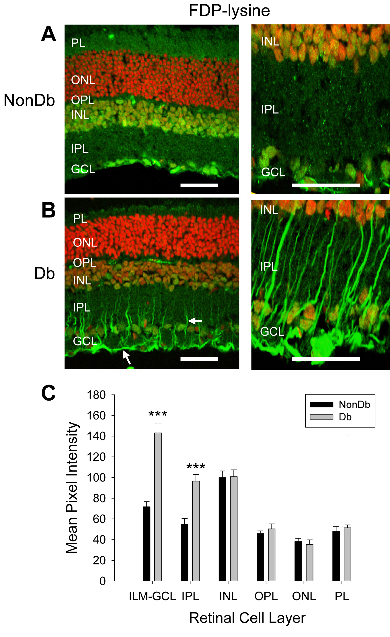
 Figure 3 of
Yong, Mol Vis 2010; 16:2524-2538.
Figure 3 of
Yong, Mol Vis 2010; 16:2524-2538.