Figure 2. Immunohistochemical staining for 4-hydroxy-2-nonenal (HNE), 4-hydroxyhexenal (HHE), and malondialdehyde (MDA) modified proteins
(green fluorescence) in transverse cryosections of retina from nondiabetic (NonDb) and diabetic (Db) rats of 4 months disease
duration. Propidium iodide was used to counterstain cell nuclei (red fluorescence). In nondiabetic retina, weak diffuse immunoreactivity
for each of these advanced lipoxidation end products (ALEs) was detected, although stronger cytoplasmic staining for 4-HHE
and MDA-modified proteins was observed for a small number of cells located within the ganglion cell layer (GCL) and at the
outer border of the inner nuclear layer (INL; arrows). Immunolabelled sections from diabetic rats appeared similar to those
of nondiabetic rats. When quantified, no significant differences in the intensity of staining for 4-HNE, 4-HHE, and MDA modified
proteins was apparent between retinas from the nondiabetic and diabetic animals.
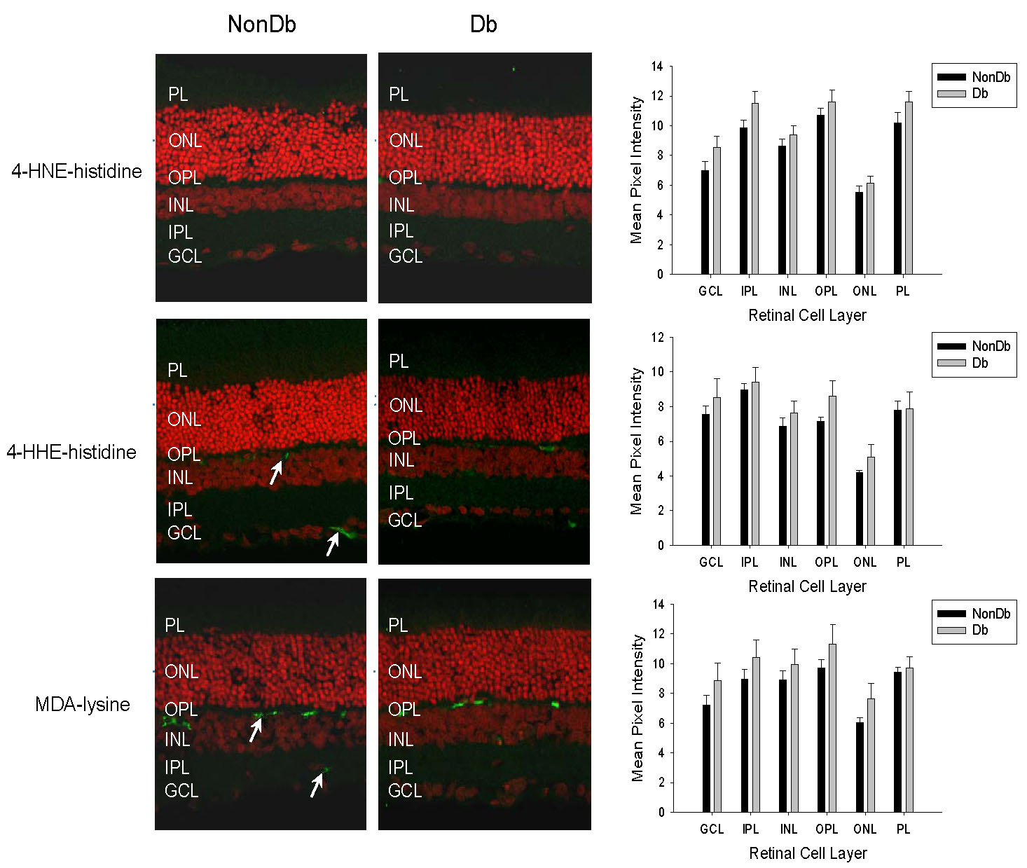
 Figure 2 of
Yong, Mol Vis 2010; 16:2524-2538.
Figure 2 of
Yong, Mol Vis 2010; 16:2524-2538.