Figure 1. Protein expression map of human trabecular meshwork by two-dimensional gel electrophoreses. Human trabecular meshwork tissue
(TMT) and cell (TMC) lysates (approximately 1 mg protein) were applied to 2-DE gel (18 cm strips) that was visualized by Coomassie
blue staining.
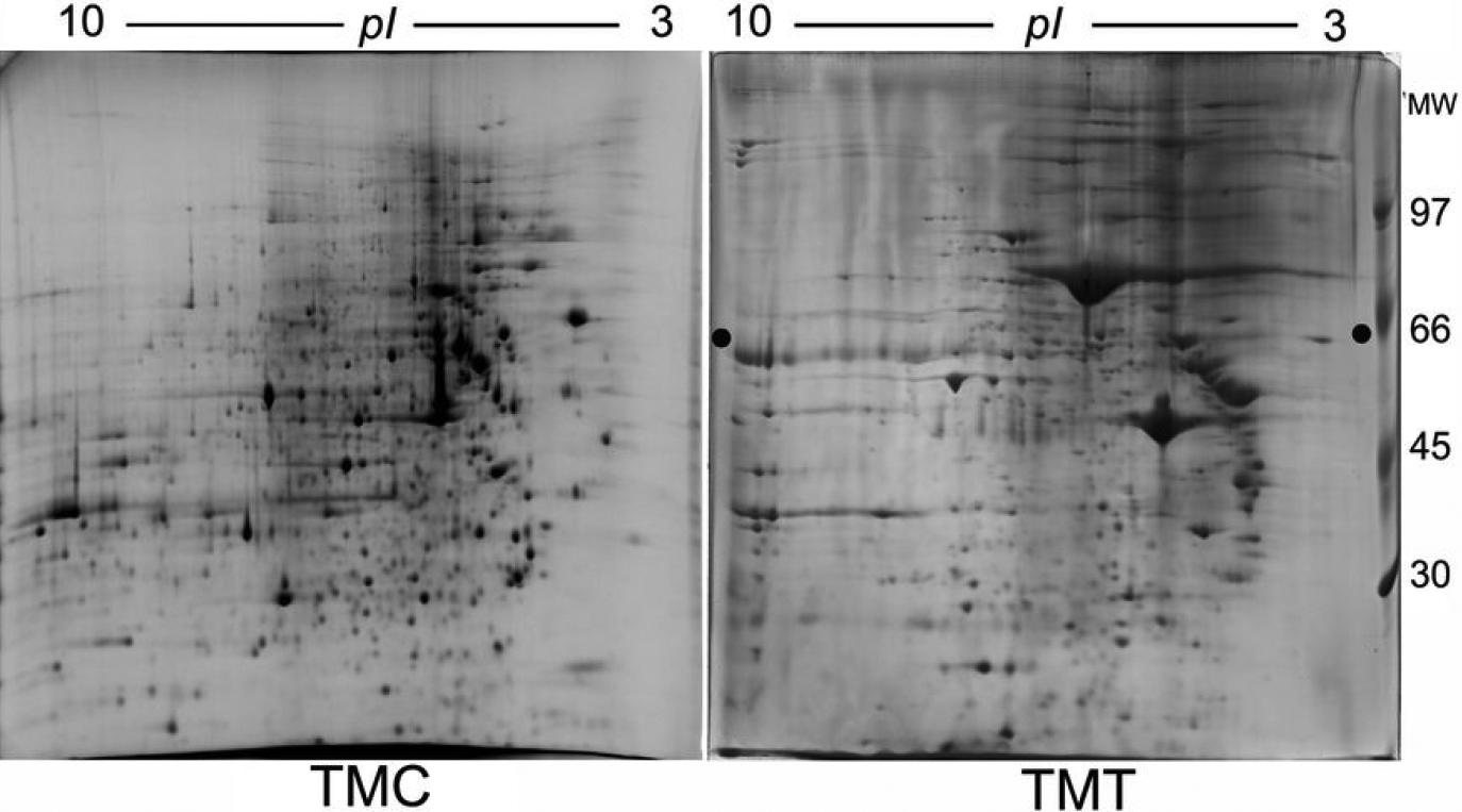
 Figure 1 of
Yu, Mol Vis 2010; 16:213-223.
Figure 1 of
Yu, Mol Vis 2010; 16:213-223.