Figure 3. Laser scanning confocal images
illustrating immunoreactivity for synemin, glial fibrillary acidic
protein, and the isolectin B4 labeling in cryosections of control
retina and following 30 days of injury. A: In control tissue,
sparse synemin immunoreactivity (red) is present in astrocytes of the
nerve fiber layer (NFL; arrows), as well as in cells along the inner
border of outer plexiform layer (OPL, asterisk), perhaps representing
the labeling of horizontal cells. B-G: After injury,
anti-synemin labeling appears as vertical streaks across the retina
representing the processes of Müller cells (B, arrows) as well
as more intense labeling of astrocytes extending processes laterally in
the NFL (B). The triple-labeled control retina shows the
presence of anti-glial fibrillary acidic protein (GFAP, green) labeled
astrocytes in the NFL, and an isolectin B4-positive blood vessel (blue,
arrow) coursing across the inner retina (C). Following long-term
injury, many Müller cells express synemin from the inner retina to the
outer limiting membrane (OLM, D-G), although this
population shows little overlap with radial processes expressing GFAP
at this time. Anti-synemin labeling also increased in astrocytes after
injury, now extending into the outer retina but still associated with
retinal blood vessels (D-G, arrows). Scale bars represent
20 μm. ONL represents outer nuclear layer; OPL represents outer
plexiform layer.
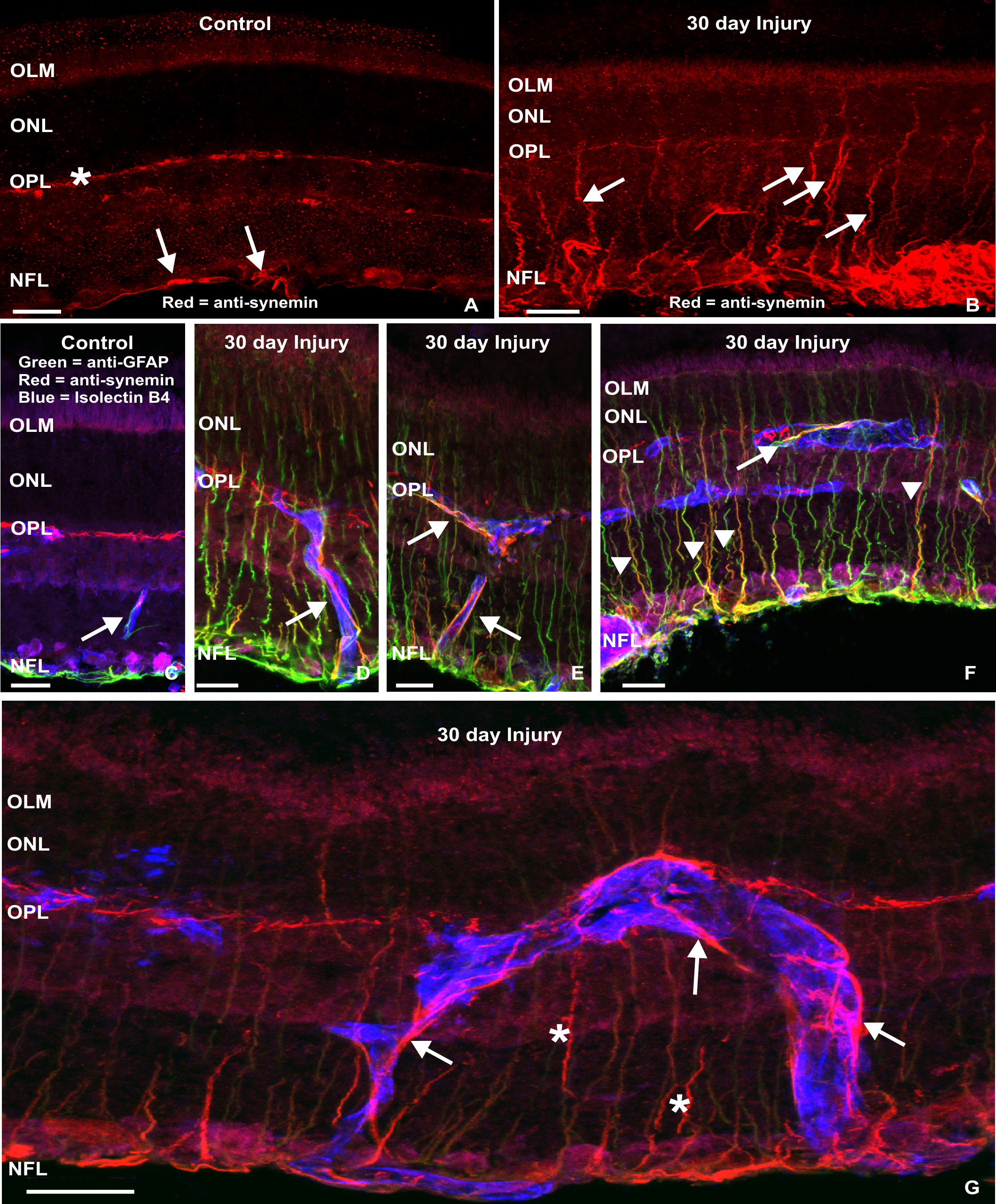
 Figure 3 of Luna, Mol Vis 2010; 16:2511-2523.