Figure 2. Laser scanning confocal images
from control and injured retinas labeled with antibodies to nestin,
bromodeoxyuridine and glial fibrillary acidic protein. A: In
control retinas, apparent anti-nestin labeling (red) due to nonspecific
binding of the anti-mouse secondary antibody (see Figure 1B) was
observed outlining blood vessels in the outer plexiform layer, while
anti-glial fibrillary acidic protein (GFAP, blue) strongly labeled
astrocytes in the nerve fiber layer (NFL). B-F: After 3
days of a sustained retinal injury, anti-nestin labeling is more
intense and more extensive in all Müller cells, but the degree of
intensity varies across the retina as shown in figures B and D.
Müller
cell nuclei that incorporate bromodeoxyuridine (BrdU, green) are
associated with areas of strong anti-nestin labeling in both figures B
and D. Anti-nestin labeling of Müller cells is strongly
associated with the presence of Müller cell outgrowth, whether at the
earliest (C, arrows), or at more advanced stages of glial scar
formation (E, D). In some cases, anti-BrdU labeled
nuclei were observed in anti-GFAP labeled cells in the NFL indicating
that astrocytic proliferation is also a component of the injury
response (D, F, asterisks). In this study anti-nestin
labeling was not observed in astrocytes. Scale bars represent 20 μm.
OLM represents outer limiting membrane; ONL represents outer nuclear
layer; OPL represents outer plexiform layer.
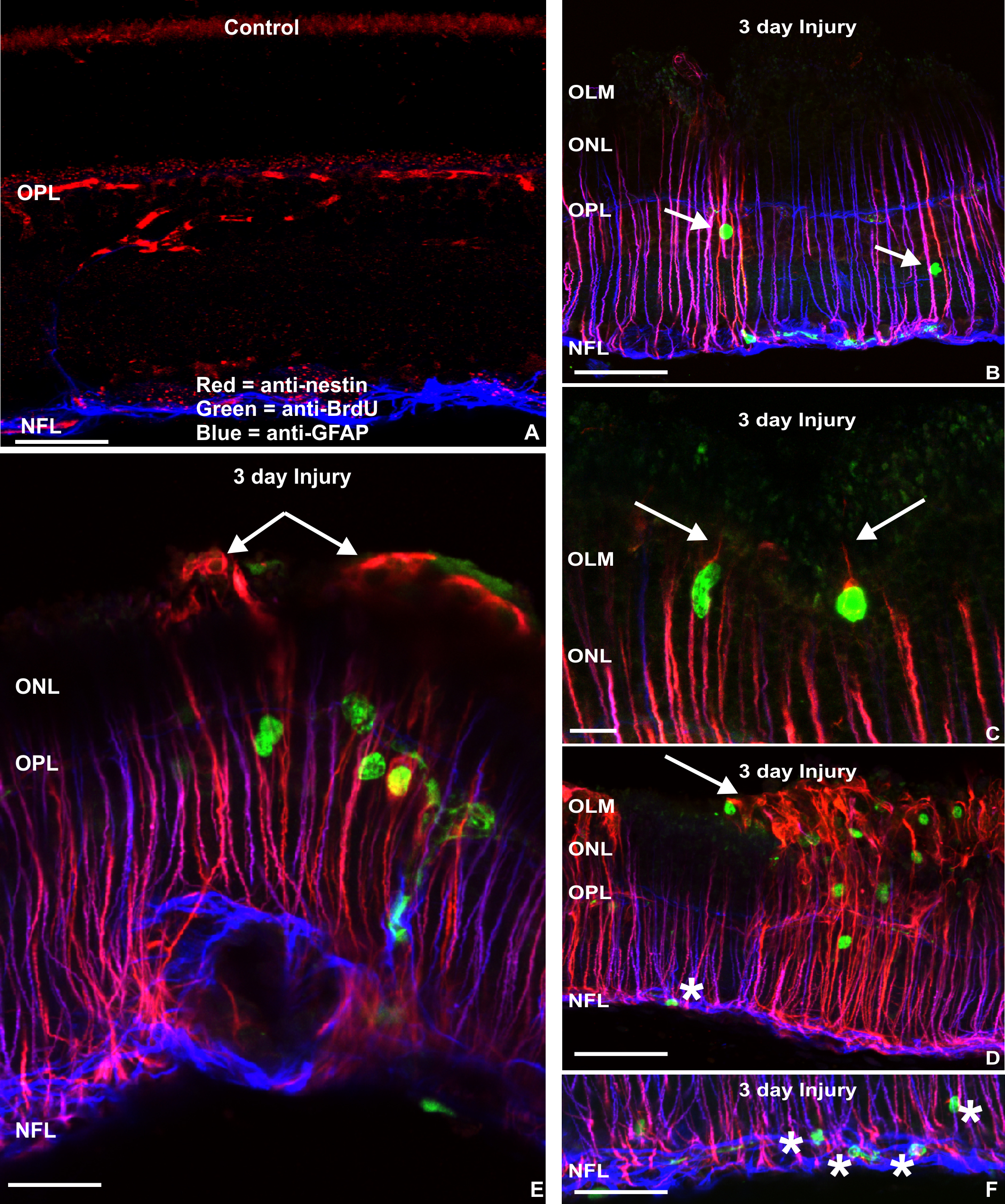
 Figure 2 of Luna, Mol Vis 2010; 16:2511-2523.