Figure 1. Laser scanning confocal images
of control and injured retinas labeled with antibodies to nestin, glial
fibrillary acidic protein, and vimentin. A, B: In the
noninjured retina, glial fibrillary acidic protein (GFAP, blue)
immunoreactivity is restricted to the very thin layer of astrocytes in
the nerve fiber layer (NFL). Apparent nestin labeling (red) in vascular
structures is due to non-specific binding of the mouse secondary IgG to
rat blood vessels as demonstrated by the secondary control data in B.
Anti-vimentin
(green) labels all of the Müller cells from the NFL into
the outer nuclear layer (ONL). C: Following 1 day of injury
these three intermediate filament proteins are greatly upregulated with
the labeling appearing as streaks extending across the retina. Note the
heterogeneity of labeling patterns among Müller cells. D-F:
Data
shown in C is divided into its three RGB channels to
demonstrate the distinctive increases in anti-nestin (E) and
anti-vimentin (F) labeling relative to anti-GFAP (D). G,
H, I: Three days following injury, the Müller cell
labeling pattern appears distinctly different from those at 1 day as
GFAP (blue) labeling increases, although the heterogeneity of
intermediate filament protein labeling remains. Strongly
nestin-positive cells resembling microglial cells in the ONL occur at
this time point (G, arrows), while Müller cells expressing
predominately nestin begin to show the formation of glial scars in the
subretinal space (I, arrow). Scale bars represent 20 μm. OPL
represents outer plexiform layer; OLM represents outer limiting
membrane.
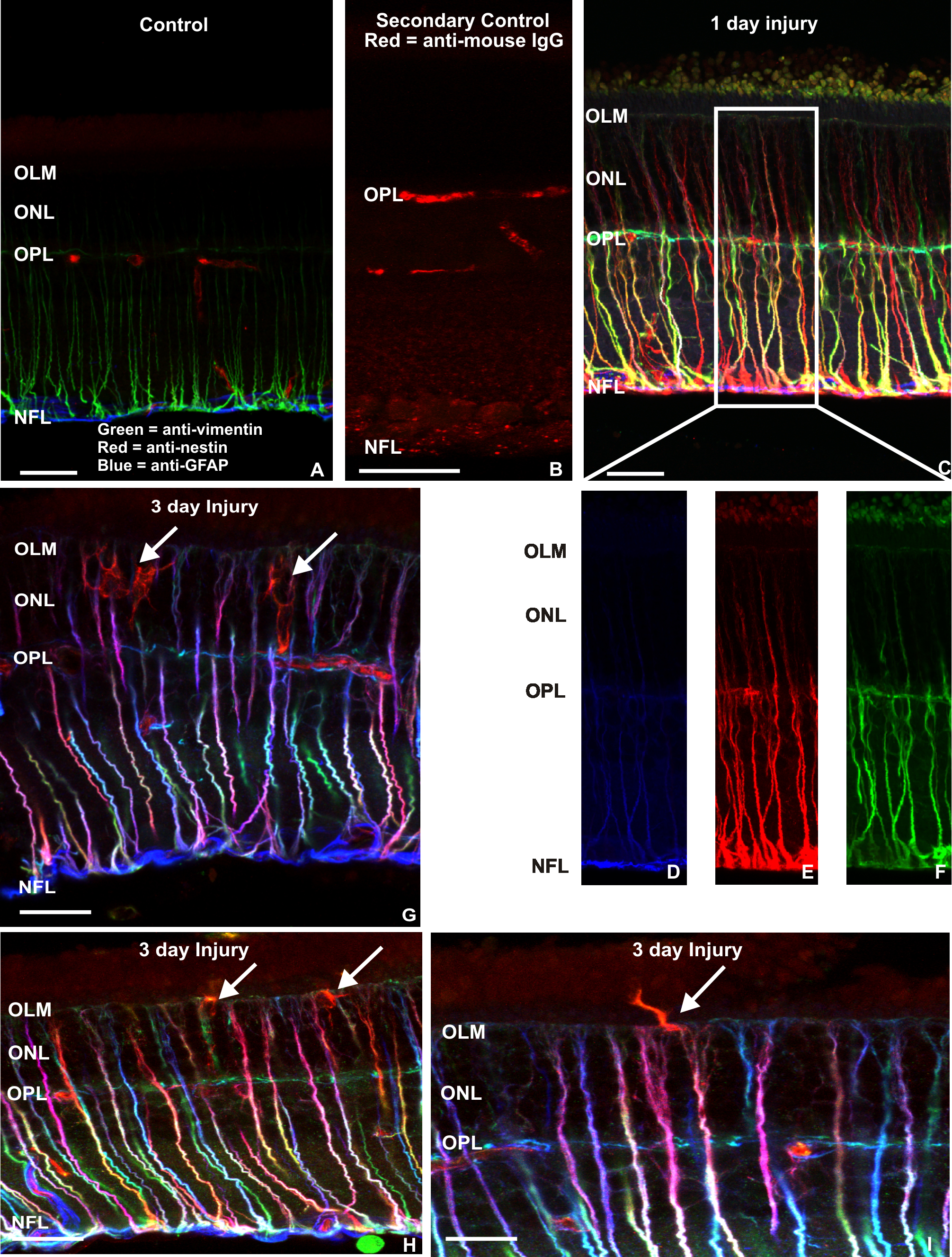
 Figure 1 of Luna, Mol Vis 2010; 16:2511-2523.