Figure 1. SV-1 can enter into culture HCE cells. A: Dose response curve of HSV-1 entry into HCE cells. Cultured HCE cells along with naturally HSV-1 resistant CHO-K1 cells
were plated in 96-well culture dishes and inoculated with twofold serial dilutions of β-galactosidase-expressing recombinant
HSV-1(KOS)gL86 virus at the plaque forming units (PFU) indicated. After 6 h, the cells were washed, permeablized and incubated
with ONPG substrate. Viral entry was measured using a spectrophotometer which measured beta-galactosidase activity at an optical
density of 410 nm. Values in the figure were plotted as the mean of three determinations (±SD). B: Confirmation of HSV-1 entry into HCE cells by X-gal staining. HCE cells grown (4×10⁶ cells) in six well dishes were inoculated with β-galactosidase-expressing HSV-1(KOS)gL86 virus at 20 PFU/cell. CHO-K1 cells
were also infected in parallel as a negative control. After 6 h of infection at 37 °C, cells were washed, fixed, and permeabilized.
X-gal was then added which yields an insoluble blue product upon hydrolysis by β-galactosidase. Blue cells, which represent
cells with viral entry, were seen in HCE cells, but not the naturally resistant CHO-K1 cells. Microscopy was performed using
a 20X objective of the Zeiss Axiovert 100 microscope. The slide book version 3.0 was used for the images.
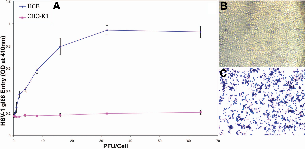
 Figure 1 of
Shah, Mol Vis 2010; 16:2476-2486.
Figure 1 of
Shah, Mol Vis 2010; 16:2476-2486.