Figure 6. Representative immunohistochemistry staining for oxidative stress and inflammatory cell markers in papillae samples from an
AKC patient. Note the presence of several inflammatory cells characterized by eosinophils in dark-pink color stained by toluidine
blue (A). The polymorph inflammatory cells are stained by the anti-CD45 antibody and evidenced by dark-brown staining (B). The presence of oxidative stress damage is observed by the immunohistochemistry staining for 4HNE (C) and HEL (D). Note that the dark-brown color represents the oxidative stress damage sites.
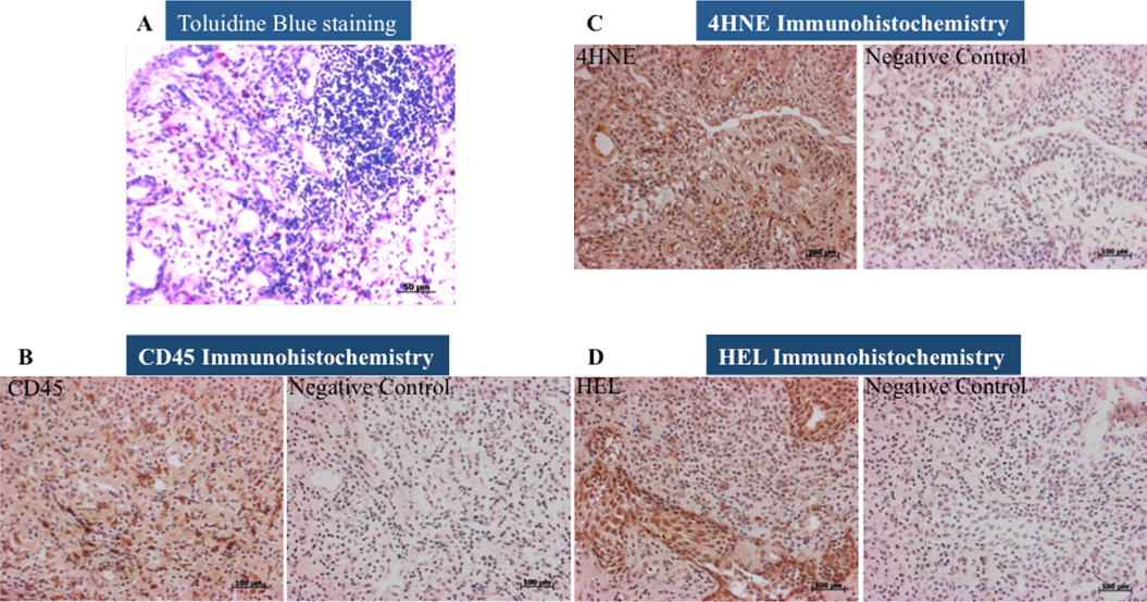
 Figure 6 of
Wakamatsu, Mol Vis 2010; 16:2465-2475.
Figure 6 of
Wakamatsu, Mol Vis 2010; 16:2465-2475.