Figure 2. Representative immunohistochemistry stainings for the early lipid oxidation marker in brush cytology specimens from an AKC
patient and a control subject. Note the extensive lipid oxidative stress damage in the HEL immunohistochemistry staining from
brush cytology samples of an AKC patient (A) compared to the healthy control subject (D). Note the significantly higher percentage of cells stained by HEL in patients with AKC (C). B and E represent the negative controls from the immunohistochemistry.
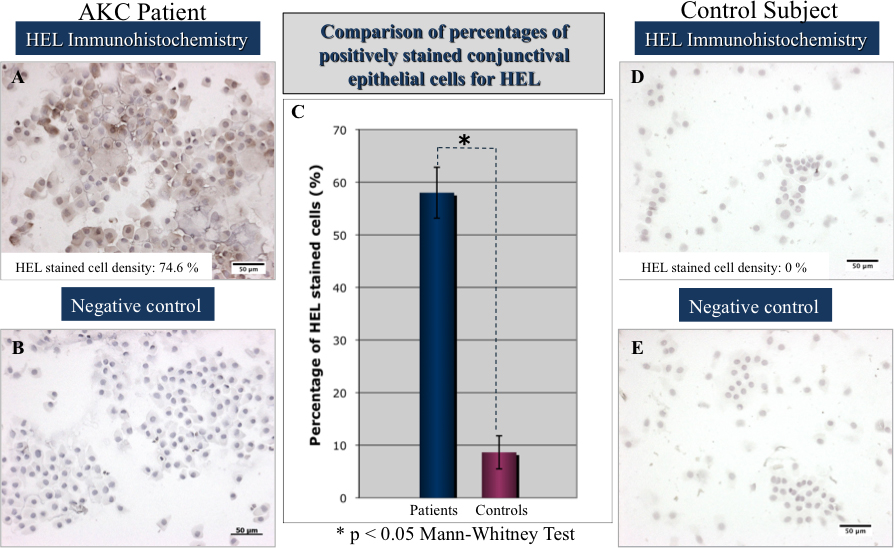
 Figure 2 of
Wakamatsu, Mol Vis 2010; 16:2465-2475.
Figure 2 of
Wakamatsu, Mol Vis 2010; 16:2465-2475.