Figure 1. Representative anterior segment photographs and brush cytology samples showing comparison of inflammatory cell infiltrates
in an AKC patient and a control. Anterior segment photographs show extensive corneal damage visualized by the fluorescein
staining in patient with AKC. Note that the superficial punctate keratopathy is present in almost all the surface of the cornea
and is associated to the increased proliferation of the conjunctival papillae (A, B). The photograph on the left side represents a normal cornea with no conjunctival proliferation on the tarsal conjunctiva
(C, D). Note the extensive inflammatory infiltrates in Diff-Quik staining from brush cytology samples in the AKC patient (E) compared with the healthy control subject (G). The graphic shows the comparison of mean percentage inflammatory cells stained by DQ between the two groups (F).
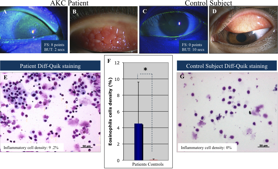
 Figure 1 of
Wakamatsu, Mol Vis 2010; 16:2465-2475.
Figure 1 of
Wakamatsu, Mol Vis 2010; 16:2465-2475.