Figure 2. Impression cytology imprint and in vivo confocal microscopy scans from a representative Sjogren syndrome patient showing nuclear
changes. A: Impression cytology imprint showing nuclear fragmentation and clumping(red arrows). B, C: Confocal microscopy scans showing these nuclear changes (red arrows).
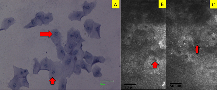
 Figure 2 of
Kojima, Mol Vis 2010; 16:2457-2464.
Figure 2 of
Kojima, Mol Vis 2010; 16:2457-2464.