Figure 6. Macrophage migration inhibitory factor enhanced mouse corneal neovascularization after suture. An 8–0 polyglactin suture was
placed in the peripheral cornea near the limbus. Seven days later, the corneal neovascularization was assayed by counting
new vessels crossing the limbus (vessel score). A-C: Representative pictures of neovascularization after MIF-incubated polyglactin suture (vessel scores: 8 [A], 6 [B], and 10 [C]). D-F: Representative pictures of neovascularization after PBS-incubated polyglactin suture (vessel scores: 6 [D], 5 [E], and 5 [F]). The violet color of the suture is visible on the corneas. G: Vessel score of the MIF group is significantly higher than that of the PBS control group (asterisk; p=0.003, Mann–Whitney
test). Error bars show mean±1.0 standard error; bars show means.
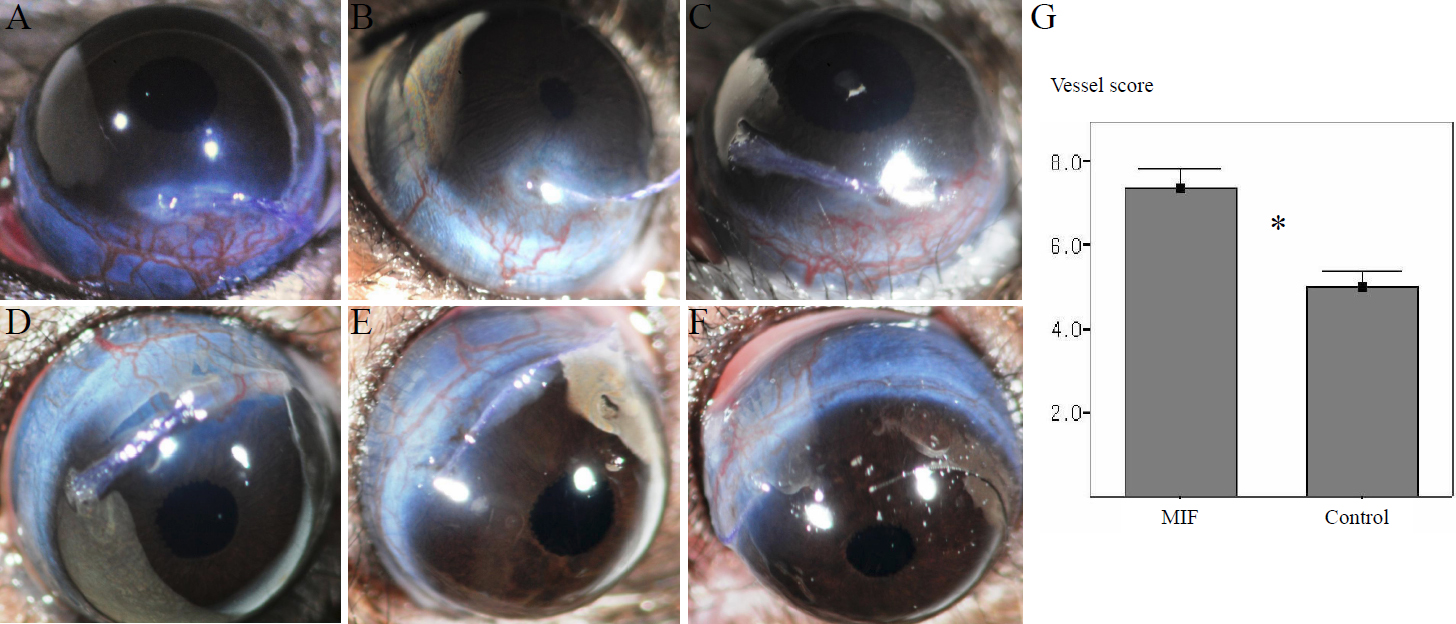
 Figure 6 of
Oh, Mol Vis 2010; 16:2402-2411.
Figure 6 of
Oh, Mol Vis 2010; 16:2402-2411.