Figure 2. Macrophage migration inhibitory
factor immunohistochemistry in normal and chemical burn applied to the
mouse eye. In normal control eyes, MIF was expressed in the corneal
epithelium, corneal endothelium, lens epithelium, and ciliary body
epithelium (A, B). However, it was difficult to find any
significant MIF staining in the corneal stroma. C: Negative
control; secondary antibody staining without primary antibody
application. D: MIF expression was detectable in corneal stroma
at day 2 after chemical burn induction. However, the infiltrating
inflammatory cells were largely negative for MIF expression (inserted
figure in panel D is the magnification of area indicated by the
clear arrow). E, F: MIF staining on days 7 and 30 after
chemical burn. MIF expression in the corneal stroma was increased but
cellular infiltration was reduced compared to day 2.
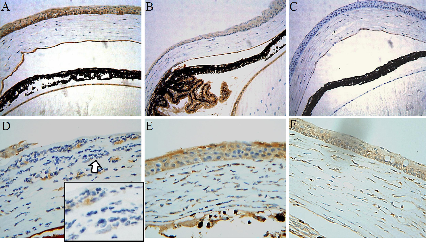
 Figure 2 of Oh, Mol Vis 2010; 16:2402-2411.