Figure 4. Endothelial regeneration. A,
B: No endothelial cells visible on the grafts, while the host
endothelium is intact. A: First day after corneal
transplantation of an endothelial cell-free syngeneic graft of group S.
B: Time of rejection in group R. C, D: Six weeks
later newly-formed endothelial cells are visible on the grafts. These
cells show an irregular shape and are larger than those in the
peripheral recipient endothelium (C: Group S; D: Group
R). A-D: Superior: Host cornea; Inferior: Graft. Black
lines: Sutures indicating the graft border. Magnification 100×. E:
Following
re-endothelialization in group S, mean endothelial cell
density on the graft is markedly lower than on the host cornea (Group
S, n=11). F: Ki-67 (MIB-1) immunostains (green) of a corneal
flatmount counterstained with DAPI (blue) shows endothelial cell
division (arrow) of host endothelium adjacent to the graft (6 days
following surgery). Magnification 630×.
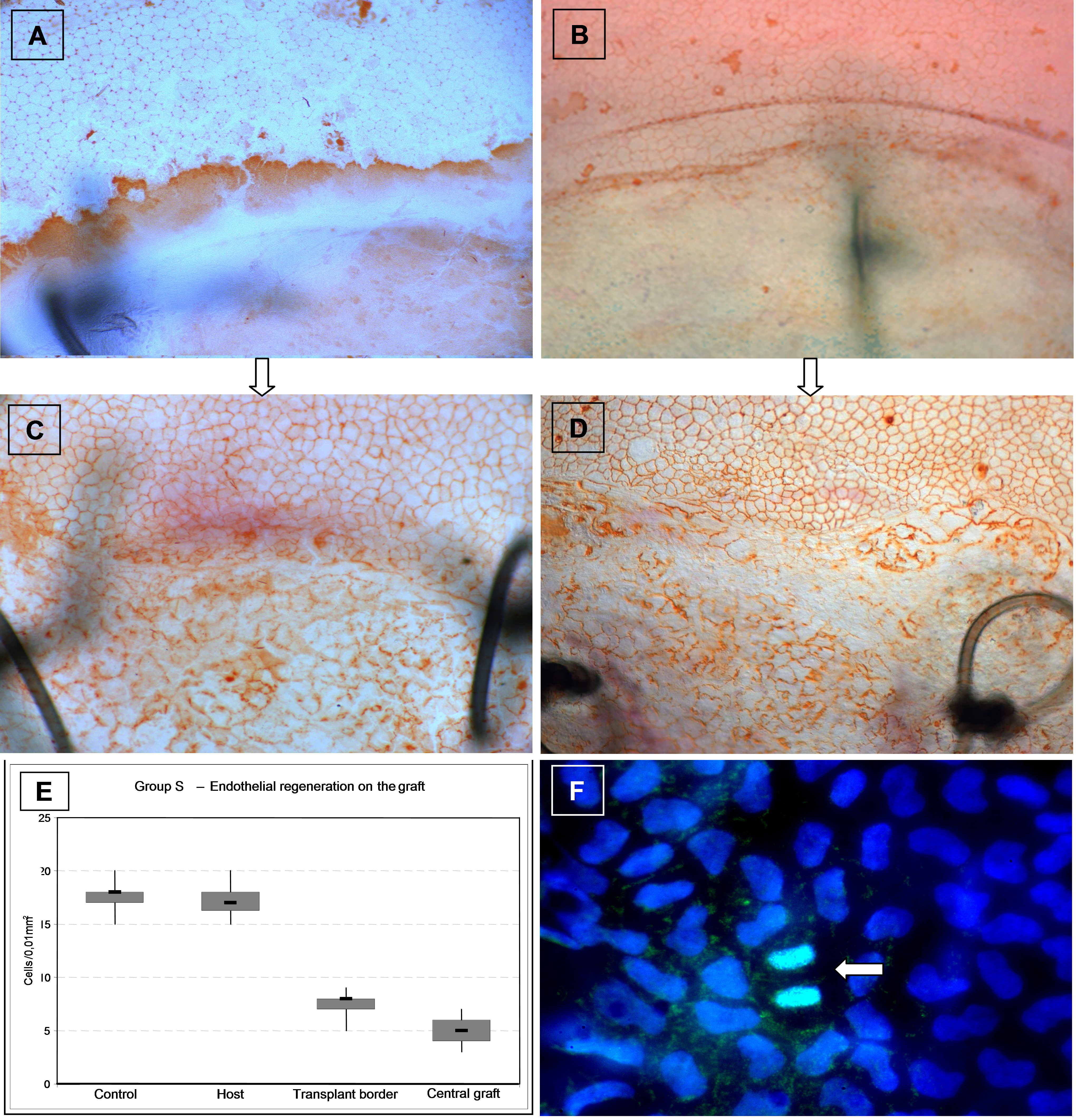
 Figure 4 of Schwartzkopff, Mol Vis 2010; 16:2368-2375.