Figure 3. Localization of hBest2 in
transfected HEK-293 cells and human ciliary body. A: HEK-293
cells were transfected with pAdlox-hBest2 and hBest2 localized using
immunofluorescence staining with GA3512 (Red in A, C, E). B:
No
hBest2 staining was observed in untransfected HEK-293 cells. C:
Although orientation varied, staining of human ciliary body identified
the basolateral plasma membrane of NPE cells. Cells in which
basolateral membrane staining is particularly evident are labeled by
white arrows. E: Staining was absent when the GA3512 was
omitted. D and F: are DIC micrographs of the same
fields shown in C and E respectively.
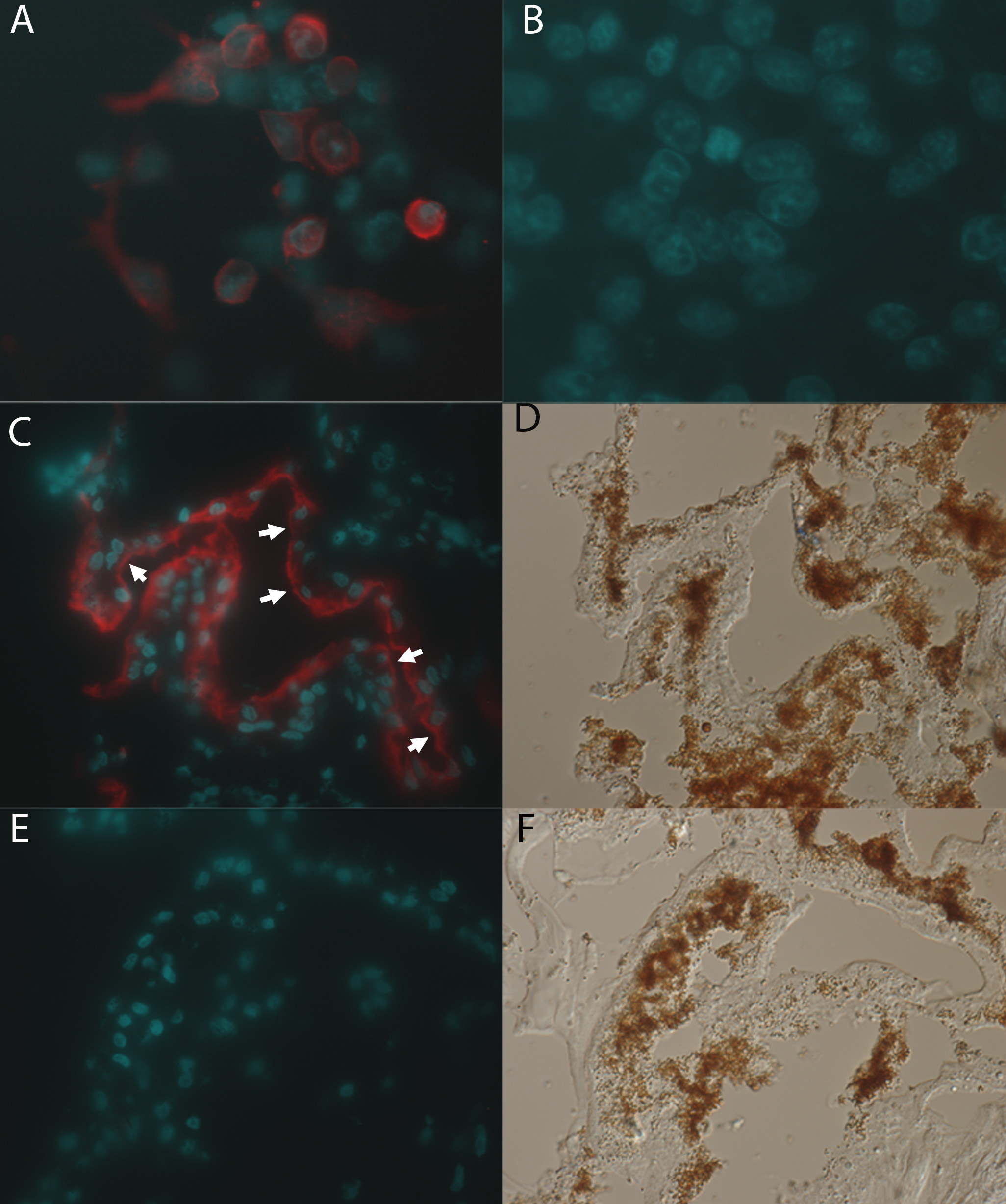
 Figure 3 of Zhang, Mol Vis 2010; 16:200-206.