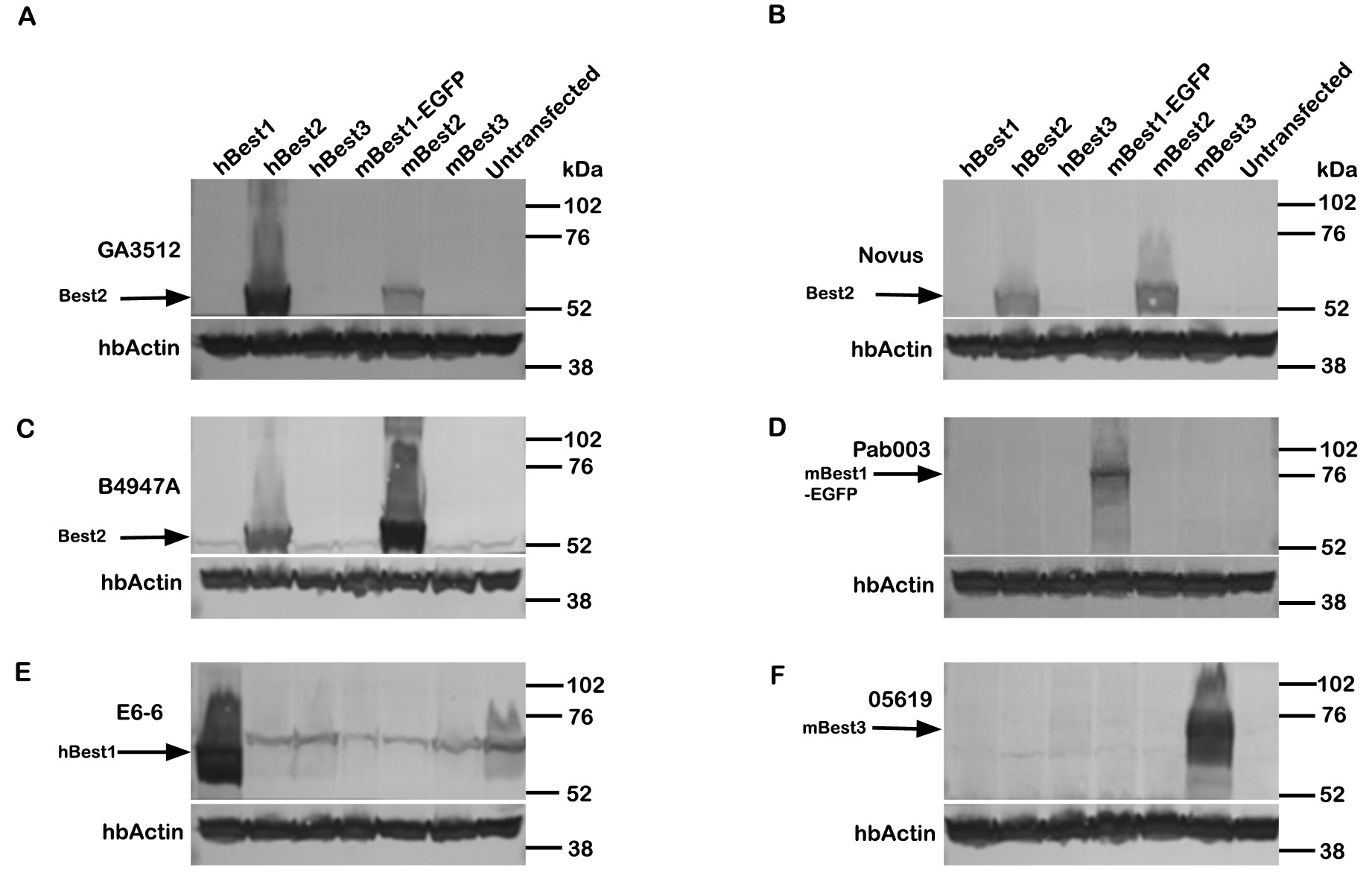
Figure 1. Specificity of GA3512. Western
blotting was performed with cell lysates from HEK293 cells transfected
to express hBest1, hBest2, hBest3, mBest1-EGFP, mBest2, or mBest3.
Untransfected HEK-293 cells were included as a negative control.
A:
GA3512
specifically
recognized
hBest2 and mBest2, but was more
efficient at identifying hBest2. It did not crossreact with other
bestrophins.
B: A commercially available anti-Best2 antibody
obtained from Novus Biologicals (NOVUS) was also specific to hbest2 and
mBest2.
C: B4947A, a rabbit polyclonal antibody against mBest2
strongly recognized mBest2 and was less effective at identifying
hBest2. Controls confirm expression of
D: hBest1,
E:
mBest1-EGFP, and
F: mBest3 using the antibodies indicated.
Interestingly, only anti-Best2 antibodies recognized both the human and
mouse forms. Also, note the approximately 70 kDa band that is
present in every lane in
E. This band is non-specific [
1].
To insure
equivalent loading, blots were cut and the bottom portion blotted for
human β-actin (hb-actin).
 Figure 1 of Zhang, Mol Vis 2010; 16:200-206.