Figure 9. A summary diagram depicting the
important structural and functional differences between the
ball-and-socket (BS) and protrusions (P) in hexagonal cortical fiber
cells. Ball-and-sockets are distributed on both the long and short
sides of fiber cells and are more frequent in the superficial than in
the deeper cortex. They are generally larger in size but smaller in
number than the protrusions distributed primarily along the corners.
Structurally, gap junctions (gj) are present in all ball-and-sockets
examined, but not in protrusions. Many elongated ball-and-socket gap
junctions protrude deeply into neighboring fiber cells. Also, while the
ball-and-socket gap junctions contain significantly different amounts
of cholesterol during fiber differentiation and maturation, all
protrusions examined consistently contain high amounts of membrane
cholesterol. The cholesterol ratio between protrusions and the
cholesterol-rich gap junctions seen in ball-and-sockets is
approximately 2:1. It is suggested that the unique structural
configuration of ball-and-socket gap junctions may significantly
facilitate cell-to-cell communication (arrows) between metabolically
active young fiber cells in the superficial cortex. Also, the large
number of ball-and-socket gap junctions found near the equatorial
region may effectively facilitate the flow of outward current toward
the equatorial surface for internal circulation of ions in the lens.
The presence of high cholesterol content in protrusions would make
these domain membranes less deformable, and would be more suitable for
maintenance of fiber-to-fiber stability during visual accommodation.
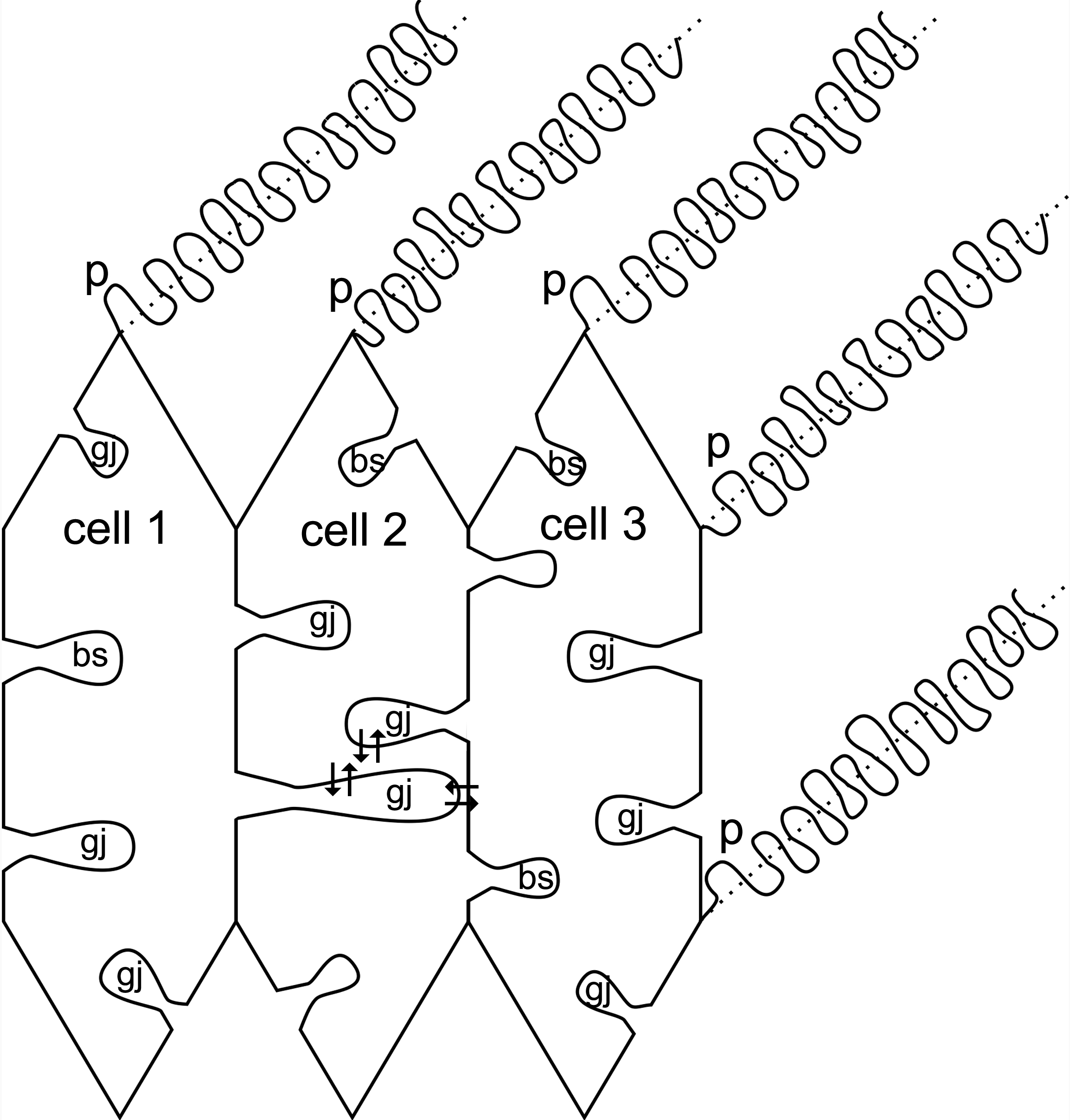
 Figure 9 of Biswas, Mol Vis 2010; 16:2328-2341.