Figure 8. Structure and cholesterol
distribution of protrusions in mature cortical fibers of the embryonic
chicken lens fibers. A: SEM showing the distribution of
numerous protrusions (arrows) from the corners of fiber cells in the
inner cortex. Several small sockets (open arrows) of ball-and-sockets
are also seen in the inner cortex. Note that two different sizes of
protrusions from adjacent cells are often paired together for
interlocking. B: Freeze-fracture TEM showing the absence of any
gap junction on protrusions (p), although two gap junctions (gj) are
found in close proximity to the protrusions (p). C:
Freeze-fracture immunogold labeling confirms the absence of labeling
for the Cx46 antibody in the protrusions (p). Instead, Cx46 antibody
specifically labels the nearby large gap junction (gj). D:
Thin-section TEM reveals the complex configuration of protrusions (p)
without the association of gap junction (open arrow) with the
protrusions. Surprisingly, several adherens junctions (paired arrows)
are found associated with the neck portion of these protrusions. E:
Filipin
cytochemistry in conjunction with freeze-fracture TEM shows
that a cluster of the protrusions (p) contain consistently high amounts
of cholesterol (filipin-cholesterol-complexes, FCCs). F: At
higher magnification, the protrusions display a high density of
membrane cholesterol (i.e., 402 FCCs/μm2 membrane). Note
that the adjacent gap junction (gj), classified as the cholesterol-rich
subtype, contains only one half of FCCs (i.e., 188 FCCs/μm2
membrane) distributed in the protrusions. G: A top-viewed
protrusion (p) in the cytoplasm showing a high density distribution of
filipin-cholesterol-complexes. The scale bar indicates 1 μm in A,
500
nm in B, C, D, E, and 200 nm in F,
and
G.
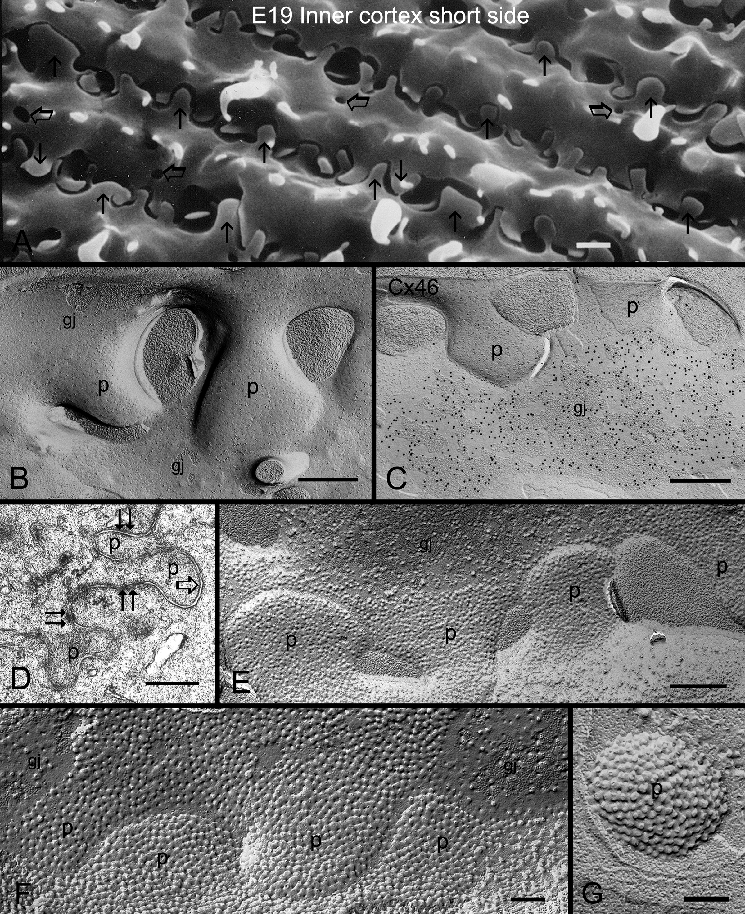
 Figure 8 of Biswas, Mol Vis 2010; 16:2328-2341.