Figure 7. Formation of ball-and-socket gap
junctions in embryonic chicken lens fibers. A: An overview of
superficial cortical fiber cells during early-stage formation of
ball-and-socket gap junctions (gj) as seen in small invaginations and
concavities (open arrows). Some small concavities (arrows) are devoid
of connexons. Others contain loosely scattered connexons which are
directly associated with nearby pools of flat membrane connexons,
suggesting that ball-and-socket connexons perhaps migrate from nearby
existing gap junctions of the flat membrane. B, C, and D:
A
representative profile showing examples of early ball-and-socket gap
junction formation. Note that differing amounts of connexons are found
distributed inside the concavities (arrow and open arrows), suggesting
that concavities are formed before the migration (or insertion) of
connexons. E: A well formed ball-and-socket domain is almost
completely occupied by gap junction connexons. The non-junctional
portion is indicated by asterisk. F and G: FRIL shows
some immunogold particles for specific labeling of Cx46 antibody are
scattered in the non-junctional portion (asterisk) inside the
ball-and-socket, suggesting the presence of individual connexons for
completion of gap junction formation in this area. The scale bars
indicate 200 nm.
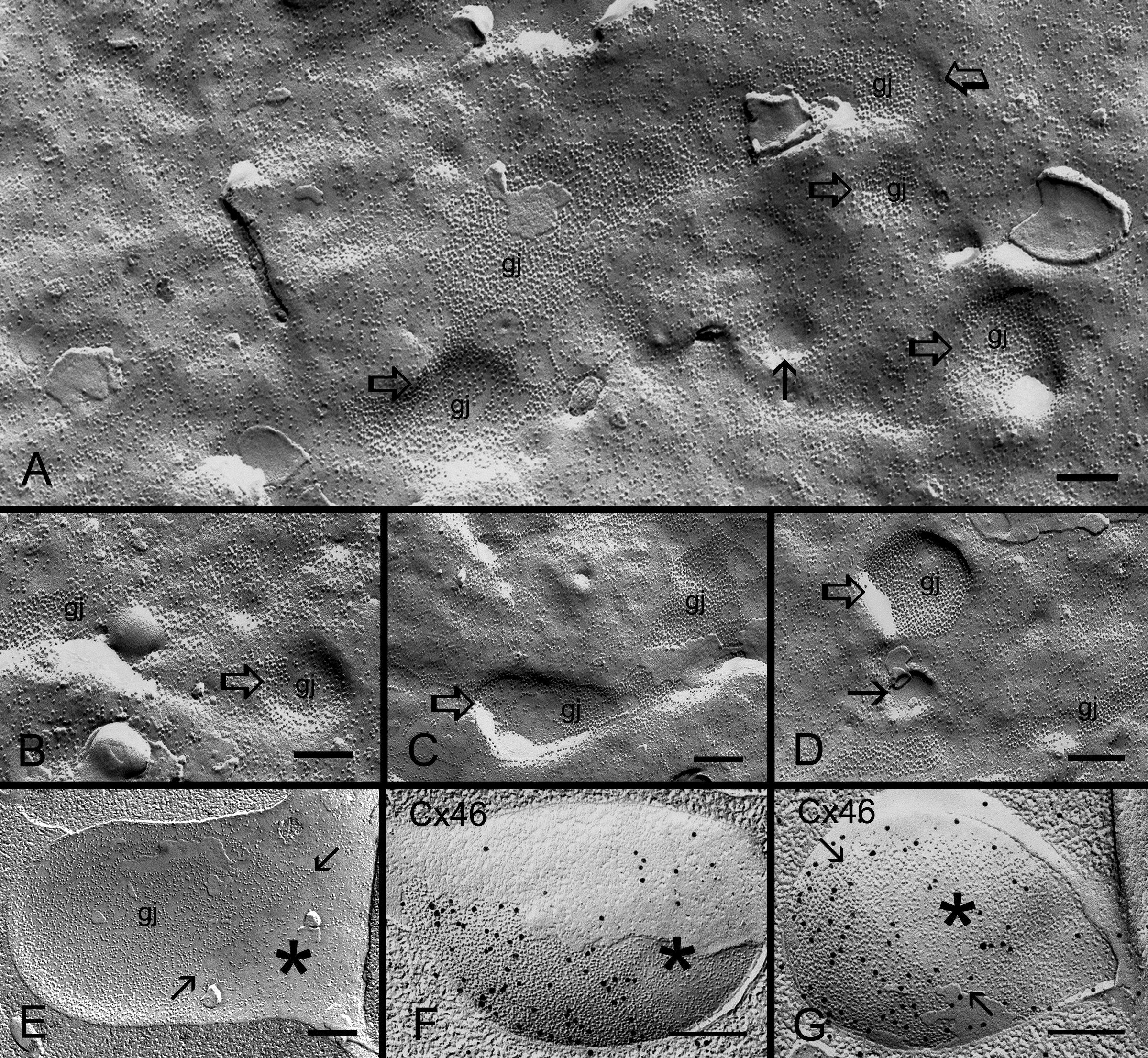
 Figure 7 of Biswas, Mol Vis 2010; 16:2328-2341.