Figure 6. Freeze-fracture TEM of
ball-and-socket gap junctions in monkey lens fibers. A: A
cluster of three shallow ball-and-socket gap junctions are seen in
superficial fibers of a young monkey (1.5 years old). B and C:
Elongated
ball-and-socket gap junctions with loosely-packed connexons
in superficial cortical fibers of a mature monkey (20 years old). Arrow
indicates the lateral cell membrane of adjacent cell. D, E,
and
F: Three different arrangements of connexons are associated
with ball-and-sockets in the deeper mature cortical fibers: (D)
A ball-and-socket completely occupied by connexons, (E) A
ball-and-socket partially occupied by connexons, and (F) A
ball-and-socket occupied by fragmentary gap junction plaques with
disorganized connexons. Ball-and-socket gap junctions in E and F
may be in a degradation stage. The scale bars indicate 200 nm.
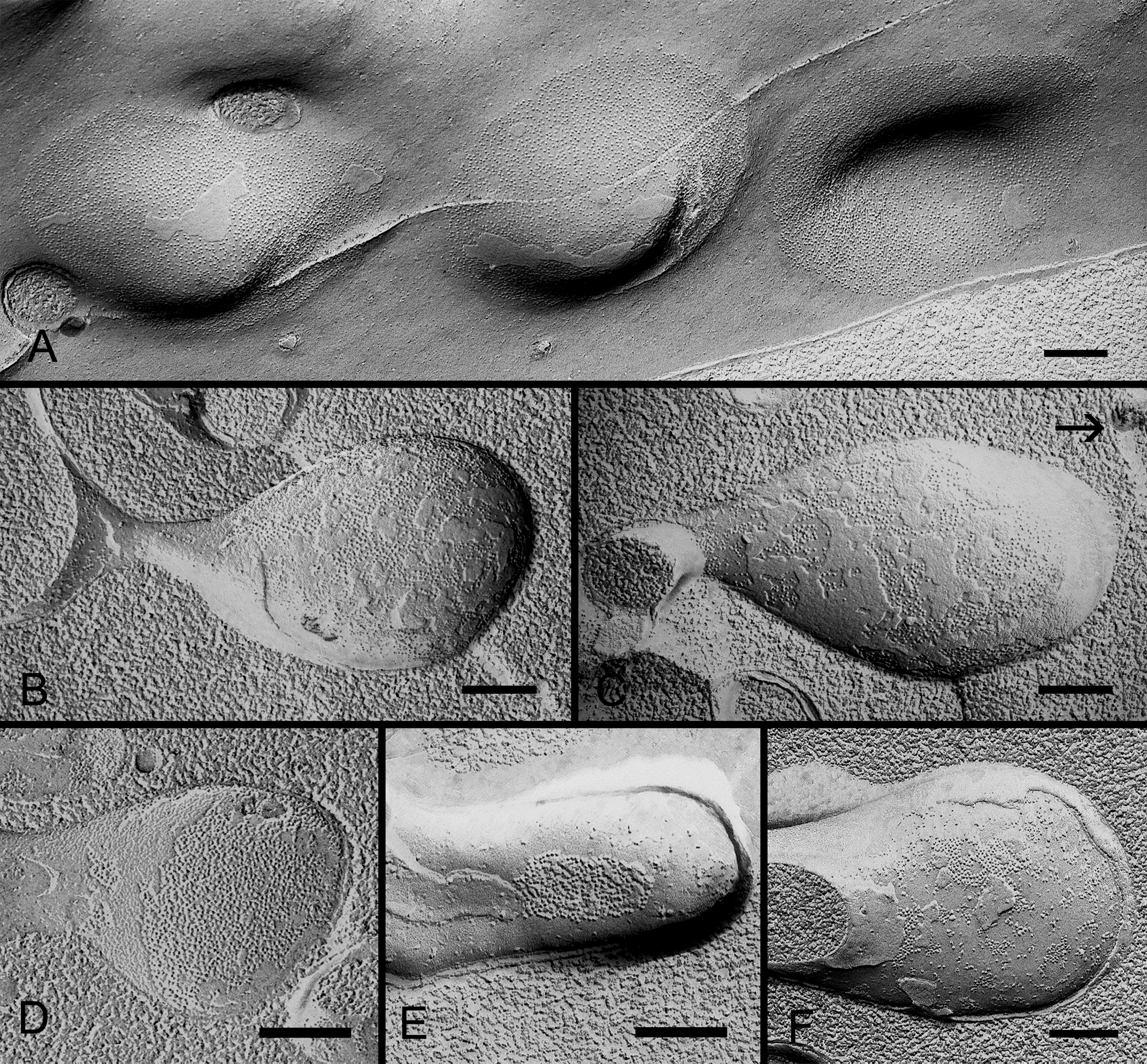
 Figure 6 of Biswas, Mol Vis 2010; 16:2328-2341.