Figure 5. Freeze-fracture TEM and
cholesterol distribution of ball-and-socket gap junctions in embryonic
and adult chicken lens fibers. A: Ball-and-socket gap junction
with loosely-packed configuration of connexons found in the outer
cortex (0–400 μm from the surface). B: Ball-and-socket gap
junction with a mixture of loosely-packed (open arrows) and
crystalline-arranged (arrows) connexons found in the deeper region of
the outer cortex. C: Ball-and-socket gap junction with
crystalline-packed connexons (arrow) found in the inner cortex (400–800
μm). D: Cholesterol-rich ball-and-socket gap junction found in
the outer cortex as determined by filipin cytochemistry in conjunction
with freeze-fracture TEM. This gap junction exhibits loosely-packed
connexons. E: Cholesterol-intermediate ball-and-socket gap
junction found in the deeper region of the outer cortex. This gap
junction displays distinct rows of crystalline-packed connexons
(arrows) and few loose connexons. F and G:
Cholesterol-free ball-and-socket gap junctions distributed in the inner
cortex. These gap junctions contain the crystalline-packed
configuration of connexons clearly seen on the P-face (pf) of the
membrane in (F) and on the E-face (ef) of the membrane in (G). H
and I: The presence of both cholesterol-rich and
cholesterol-poor gap junctions in ball-and-sockets of the embryonic
lens at E20. Several filipin-cholesterol-complexes (arrows) are
indicated in the ball-and-socket in (I). The scale bars indicate
200 nm.
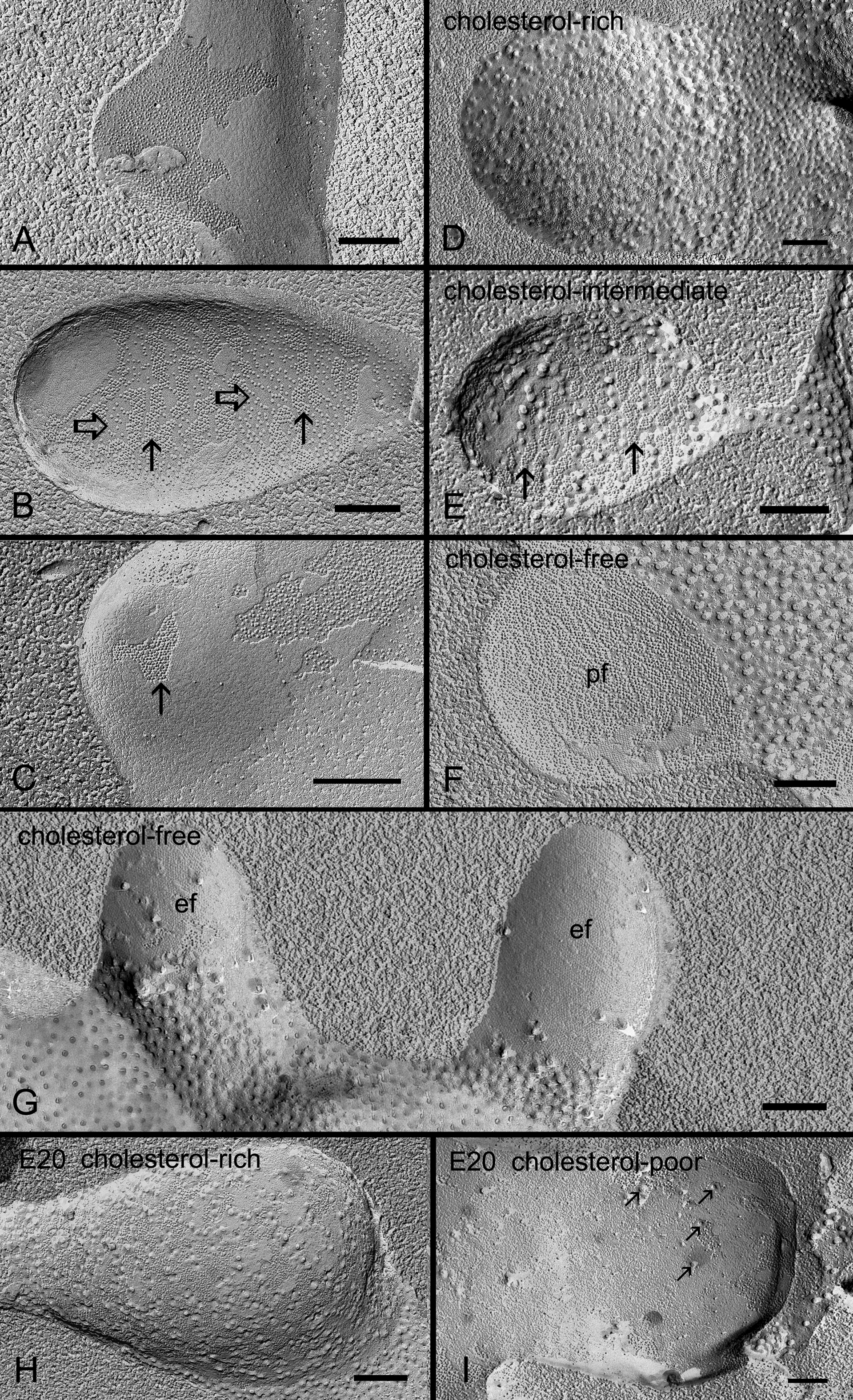
 Figure 5 of Biswas, Mol Vis 2010; 16:2328-2341.