Figure 4. Freeze-fracture TEM and
freeze-fracture immunogold labeling of Cx46 and Cx50 in ball-and-socket
gap junctions of embryonic chicken lens fibers. A:
Freeze-fracture TEM showing distribution of a cluster of
ball-and-sockets containing gap junctions (gj). B: Higher
magnification reveals the presence of gap junction plaque in the entire
ball-and-socket domain (from cell 1) which protrudes into the cytoplasm
of cell 2. The tip of this gap junction is in close proximity to the
cell membrane of cell 3 which contains several flat gap junctions (gj).
C: An elongated ball-and-socket gap junction, ~3.5 μm long and
~1.8 μm wide, from cell 1 protrudes deeply into the neighboring cell 2,
and almost makes direct contact with the lateral cell membrane (open
arrow) of cell 3. D: High magnification from the area marked by
arrow in C showing gap junction particles (connexons) clearly
seen on the P-face (pf) of the junctional membrane. E and F:
FRIL
shows the specific labeling of Cx46 and Cx50 antibodies in the
ball-and-socket gap junctions, respectively. The scale bar indicates 1
μm in A; 500 nm in B and C; 100 nm in D,
and
200 nm in E and F.
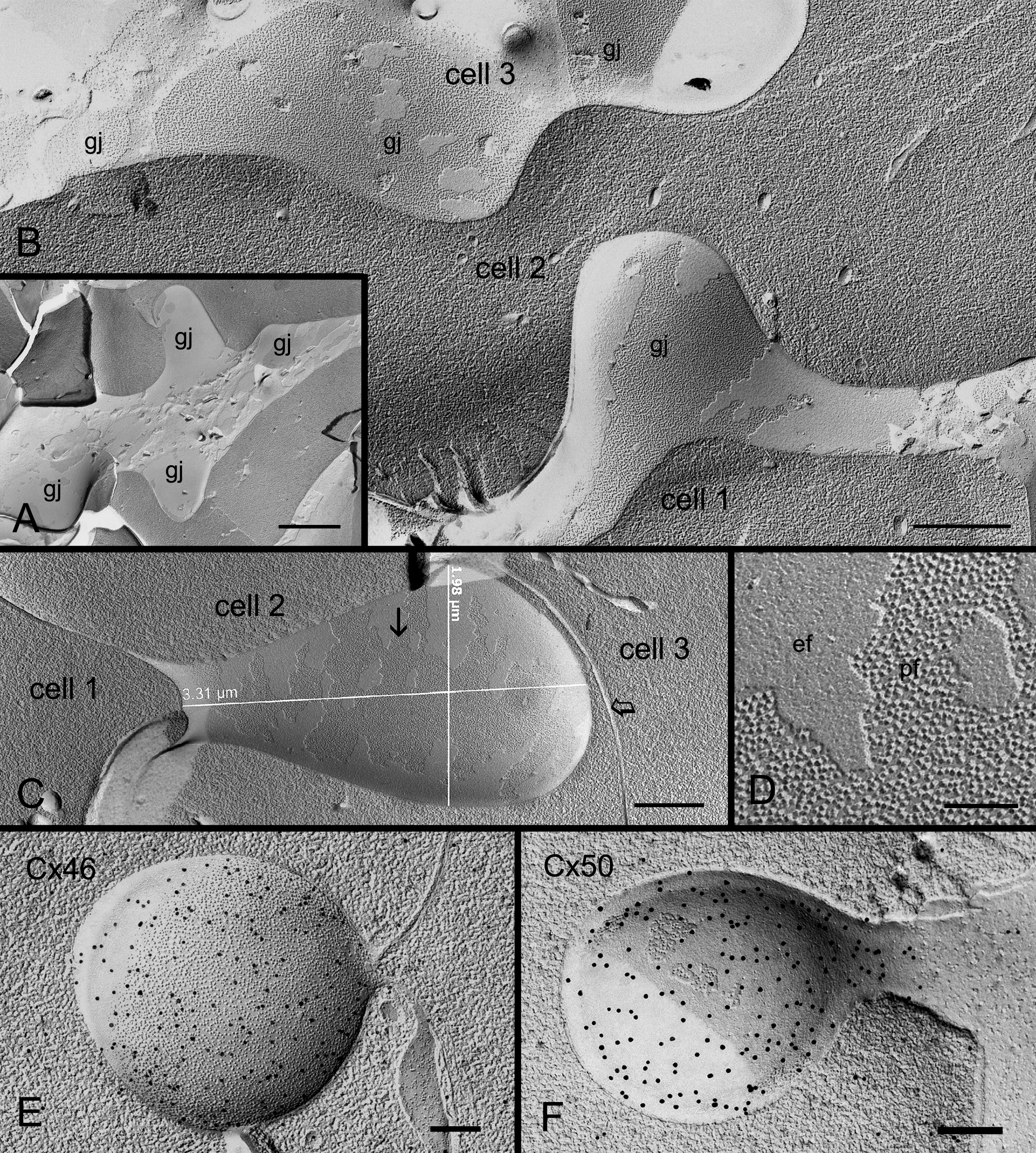
 Figure 4 of Biswas, Mol Vis 2010; 16:2328-2341.