Figure 3. SEM of ball-and-sockets in
different cortical regions of monkey lens (20 year old). A:
Superficial cortical fibers (approximately 100 μm from the surface),
numerous ball-and-sockets are distributed on the long side of fiber
cells. B: Intermediate cortical fibers (approximately 300 μm
from the surface), a large number of ball-and-sockets are found on the
long side of fiber cells. In this region, many protrusions (p) are also
distributed along the corners of cortical fiber cells. C:
However, in the deeper cortex (approximately 500 μm from the surface),
ball-and-sockets display smaller number and size with degenerating
appearance. The scale bars indicate 1 μm.
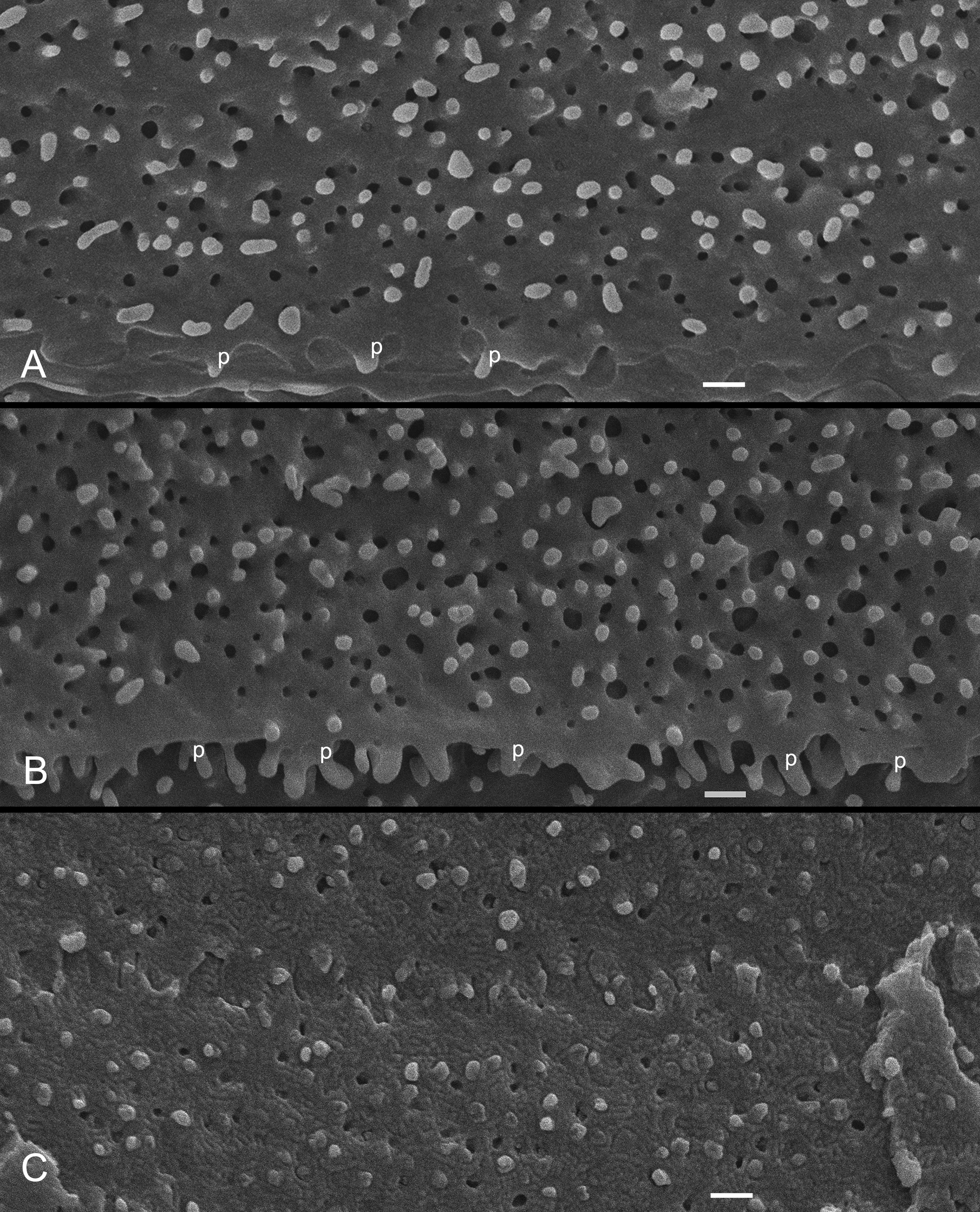
 Figure 3 of Biswas, Mol Vis 2010; 16:2328-2341.