Figure 2. SEM and thin-section TEM of
ball-and-sockets in the embryonic and adult chicken lenses. A:
In the embryonic chicken lens (E18), a representative scanning electron
micrograph showing many ball-and-sockets distributed on the short sides
of superficial cortical fibers at the equatorial region (approximately
100 μm from the surface). B: At higher magnification, the
height and shape of the ball-and-sockets can be readily visualized from
side-view. C: In the adult chicken lens (P42 weeks), numerous
ball-and-sockets are seen distributed in rows on the two long sides of
superficial cortical fibers at the equatorial region (approximately 200
μm from the surface). It is estimated that the number of
ball-and-sockets (approximately 13 ball-and-sockets per 100 μm2
membrane) is not significantly different at the apical, equatorial and
posterior regions along the anterior-posterior axis of any given
superficial fiber cell. The border between two long-side fiber cells is
marked by two arrows. Note that the height and the shape of these
top-viewed ball-and-sockets cannot be readily appreciated as compared
with those seen from the short sides in A and B. D:
Thin-section
TEM shows a ball-and-socket with the head and neck
portions protruding into an adjacent cell. This ball-and-socket is
completely occupied by a gap junction (gj). E: A cross-section
of the ball-and-socket gap junction showing a considerable membrane
extension of this junction into a narrow fiber cell. In the images, b
is the ball, and s is the socket. The scale bar indicates 5 μm in A,
C, 2 μm in B, and 200 nm in D and E.
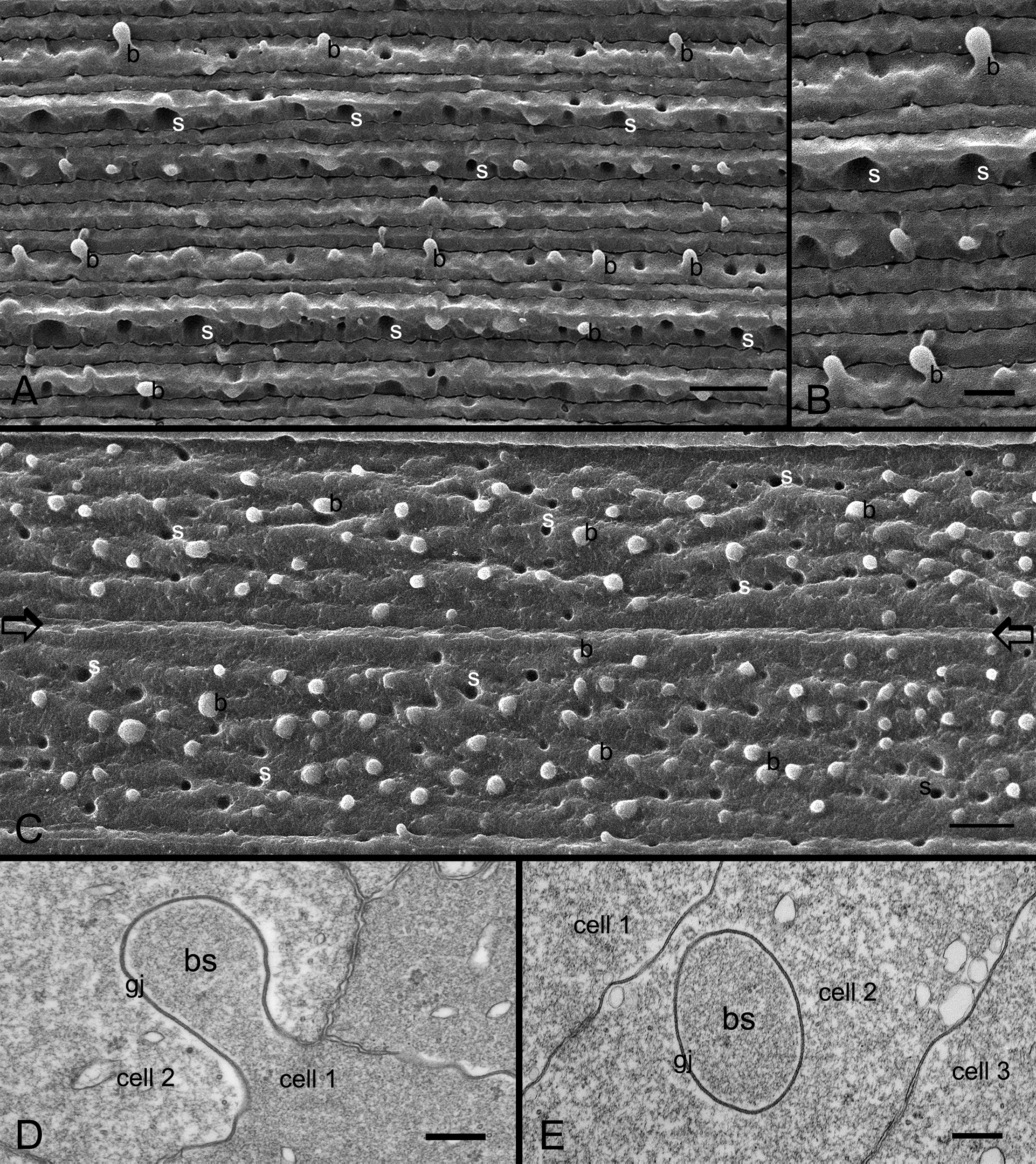
 Figure 2 of Biswas, Mol Vis 2010; 16:2328-2341.